


A 56-year-old patient admitted due to cardiogenic shock following an acute myocardial infarction. Peripheral veno-arterial extracorporeal oxygenation membrane (VA ECMO) was implanted as a bridge to decision. Five days later, patient developed differential hypoxemia between upper (SO2 77%) and lower body (SO2 100%). An echocardiogram showed left ventricular ejection fraction improvement from 10% to 30% and pulmonary ultrasound revealed a mild pleural effusion and a bilateral image of pulmonary consolidation with dynamic air bronchogram. Inside the bronchus, vermiform mobile hyperechoic images (black arrows, figure 1A) appeared synchronized with breathing (video 1). Simultaneous bronchoscopy showed purulent secretions (white arrow, figure 1B) going in and out of the bronchial tree (video 2). In patients with VA ECMO, beyond the echocardiogram, pulmonary ultrasound is essential to see lung patterns that could explain hypoxic and even to guide therapeutic attitudes.
Conflict of Interest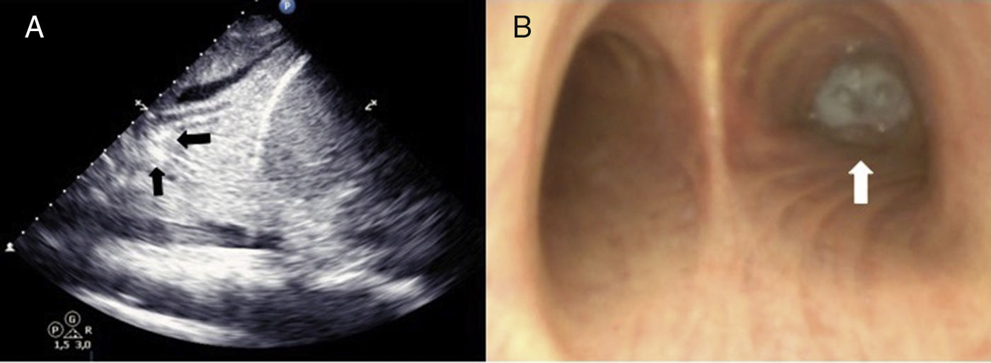
The authors declare no conflicts of interest.

