
Central venous cannulation (CVC) is common and necessary in pediatric intensive care. However, this procedure is not without risks or complications. Although CVCs have classically been placed following anatomical landmarks, the use of ultrasound guidance has largely replaced the latter, given its better profile of efficacy and safety, demonstrated at least in adult populations.
ObjectivesTo compare the effectiveness and safety in the insertion of femoral central venous catheters guided by ultrasound (US) versus the anatomical method (LM) in critical care pediatric patients.
Methods100 patients were randomized: 50 were assigned to the US group and 49 to the LM group. In the LM group the traditional method consisted in palpating the femoral artery pulse as a; in the US group the CVC was inserted using a real time technique. Success at the first attempt, overall success in cannulation, number of attempts and arterial puncture were the variables studied in both groups.
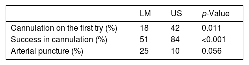
ResultsSuccess at the first attempt and overall success in cannulation were significantly higher in the US group versus the LM (US 42% vs. LM 18%, p 0.011, US 84% vs. LM 51% p <0.001, respectively). The incidence of puncture of the femoral artery was lower in the US group (LM 12 vs. US 5, p 0.056) without achieving statistical significance.
ConclusionsAccording to our results, the placement of central venous access via the femoral approach should be preferably performed under ultrasound guidance, however, further studies in larger populations are needed to confirm this findings.
La instalación de un catéter venoso central (CVC) es una práctica común y necesaria en cuidados intensivos pediátricos. Sin embargo, dicho procedimiento no se encuentra carente de riesgos ni de complicaciones. Si bien clásicamente los CVC se han colocado siguiendo referencias anatómicas, el uso de la guía ecográfica ha reemplazado a este método, dado su mejor perfil de eficacia y seguridad, demostrado al menos en poblaciones adultas.
ObjetivosComparar la efectividad y seguridad en la inserción de catéteres venosos centrales femorales guiados por ecografía (US) versus mediante el método anatómico (LM) en pacientes críticos pediátricos.
MétodosSe aleatorizaron 100 pacientes: 50 se destinaron al grupo US y 49 al LM. En el grupo LM se usó el método tradicional palpando pulso femoral; en el grupo US se colocó el CVC guiado por ecografía en tiempo real. El éxito al primer intento, éxito global en la canulación, número de intentos y la punción arterial fueron las variables de estudio entre ambos grupos.
ResultadosEl éxito al primer intento y el éxito global en la canulación fueron significativamente mayores en el grupo US versus LM (US 42% vs. LM 18%, p=0,011; US 84% vs. LM 51%, p<0,001, respectivamente). La incidencia de punción de la arteria femoral fue menor en el grupo US (LM 12 vs. US 5, p=0,056) sin lograr significación estadística.
ConclusionesDe acuerdo con nuestros resultados, la colocación del acceso venoso central por vía femoral debiera realizarse bajo guía ecográfica, aunque se necesitan estudios en poblaciones más numerosas que confirmen dichos resultados.
Central venous cannulation is common and necessary in pediatric intensive care. However, this procedure is not without risks or complications. Although CVCs have classically been placed following anatomical landmarks, the use of ultrasound guidance has largely replaced the latter, given its better profile of efficacy and safety, clearly demonstrated at least in adult populations.1,2
The use of ultrasound is well-known and respected, however, there are not large well-designed studies in this setting and there is more evidence in adults compared with pediatric population.3–5 In critically-ill pediatric patients data shows advantage of ultrasound guidance in the insertion of central catheters via the internal jugular vein access5–7; regarding femoral approach, there are published two randomized studies8,9 comparing the landmark technique vs. the ultrasounds guidance, with inconclusive evidence for supporting one method over the other.
As part of the quality search process in patient care and given the lack of evidence in this area, we designed a randomized multicenter study in the pediatric critical care unit (PICU) comparing the landmark (LM) with real-time ultrasound guidance (US) for inserting central venous catheters via the femoral approach. Our primary outcome is the cannulation rate at the first attempt of the CVC in the femoral vein, and secondary outcomes include successful insertion, number of attempts and incidence of femoral artery puncture.
This work is registered in ClinicalTrials.gov identifier NTC02318940. Available at: https://clinicaltrials.gov/ct2/show/NTC02318940?term=02318940; in addition, it has financing of the Research Unit, Luis Calvo Mackenna's Hospital, and East Department of the University of Chile.
MethodStudy designMulticenter randomized prospective study in the PICUs of the “Roberto Del Río” and “Exequiel González Cortés” Children's Hospitals. Patients entered the study between February 2015 and November 2015, after signing their informed consent by their parents or legal representative. This study was reviewed and approved by the local institutional review board (IRB).
PatientsAll the newly admitted patients who met criteria for admission to the PICU and who needed a central venous access according to the criteria of treating physician were enrolled in this study. Children younger than 7 days and older than 15 years, those had local infection at the puncture site, known anatomical and/or functional vascular alterations were excluded.
Subgroup analysis will be considered a priori in patients younger than 6 months.
Sample sizePrevious studies report an average success rate of 50% at the first attempt using the LM method, both in children and adults.9–11 Sample size is determined to improve the success rate by 30% using US guidance. To obtain a power of 80% in relation to success at the first attempt and with a confidence level of 95%, a number of 50 patients per branch was determined.
Randomization methodRandomization was performed by blocks for each center, with the Stata® 12.0 Software. They were stored in sealed envelopes available 24h a day in the unit. The selection of the vein was according to the attending physician, and patients selected for femoral CVC insertion were included consecutively.
OperatorThe insertion, LM and US, was performed by a pediatric intensivist or by a resident of the pediatric intensive care university program at the PICUs of the hospitals involved in this study.
To standardize knowledge in the placement of the CVC, intensivists were trained in ultrasound-guided cannulation by an expert pediatric intensivist, consisting in 2h of theory time and two hours of practice time: this was performed in phantoms specially designed for pediatric patients with small vessels and using Mindray® ultrasound machines.
Insertion method12- -
LM technique: In the supine position with external rotation and abduction of the lower extremity, the femoral artery is located by palpation in the femoral triangle and is punctured medially in the direction of the navel until it has reflux of venous blood.
- -
US Guidance: Ultrasound was performed to verify the patency and position of the target vessel before cannulation, which was performed using and in-plane or an out-plane technique (based on operator preference). The transductor and cable were covered by sterile material.
- -
Successful cannulation: it was considered when the catheter is placed without difficulty in the femoral vein.
- -
Cannulation on the first attempt: it was considered when the CVC insertion is achieved at the first transcutaneous passage to the needle.
- -
Attempt of cannulation: it was considered an attempt to pass the needle without withdrawing or redirecting the needle with forward movement. Each successive removal or redirection with a forward movement was considered one separate attempt.
- -
Arterial puncture: The arterial puncture involves aspiration of pulsatile arterial blood for both methods, and also observing the needle in the artery in the US method.
- -
Rescue: After the fifth failed attempt in LM group, up to five attempts with ultrasound guidance were made in the same vein.
- -
US machine: For this study, a Sonosite® M-Turbo (Bothell, USA) equipped with a “7–10MHz” linear transducer (“Roberto del Río” Hospital) and a General Electric Vivid q (Bothell, USA) equipped with a 12MHz linear probe (“Exequiel Gonzales Cortes” Hospital) were used. Both US machines have software to improve the display of the needle.
- -
Catheter: the catheter was chosen based on patient size; 4 and 5F and 8cm length catheters were the most used.
The main outcome was the insertion of the catheter at the first attempt; secondary outcome was the overall success in cannulation, number of attempts and, as a complication of the procedure, the rate of arterial puncture.
Rescue was used after the 5th attempt in LM technique, changing to US Guidance.
Failure to achieve access in both methods was considered as unsuccessful cannulation and the puncture site was change.
Statistical analysisDescriptive analysis was performed for continuous variables with normal distribution using means and standard deviations, and continuous variables of not normal distribution and qualitative variables with medians and percentiles. For the primary outcome, proportions are compared for dichotomous variables using the Chi-square method. A value of p <0.05 will be considered significant. Stata® 12.0 Software will be used for statistical analysis.
Multivariate Logistic Regression with Stepwise technique is performed in order to recognize independence of the method used in Outcome versus other variables that could be influencing the final result.
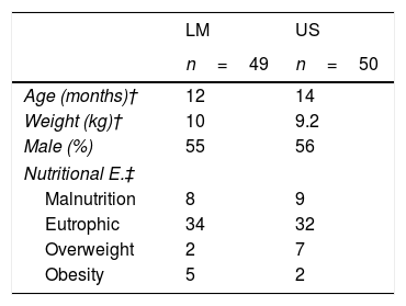
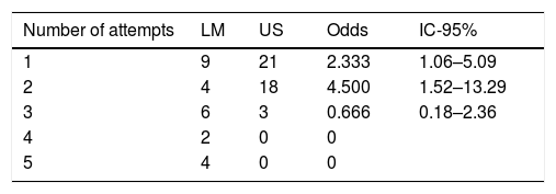
ResultsNinety-nine patients were recruited, 50 in the US group and 49 in the LM group, one patient was lost during randomization. The characteristics of the patients were similar for both groups (Table 1). The success on the insertion of the catheter at the first attempt was significantly higher in the US group (US 42% vs. LM 18%, p 0.011) as well as overall cannulation success (US 84% vs. LM51% p <0.001) (Table 2). In relation to the number of attempts to achieve successful cannulation, again the ultrasound method was superior to the LM technique (Table 3). While the protocol includes rescues, few of them were performed and thus a more thorough analysis cannot be performed
Patient characteristics.
| LM | US | |
|---|---|---|
| n=49 | n=50 | |
| Age (months)† | 12 | 14 |
| Weight (kg)† | 10 | 9.2 |
| Male (%) | 55 | 56 |
| Nutritional E.‡ | ||
| Malnutrition | 8 | 9 |
| Eutrophic | 34 | 32 |
| Overweight | 2 | 7 |
| Obesity | 5 | 2 |
LM, landmark method.
US real-time ultrasound guidance method.
Data expressed in medians † and numbers ‡.
There was no statistical difference between the two groups. Considering significant p-value <0.05.
Number of attempts.
| Number of attempts | LM | US | Odds | IC-95% |
|---|---|---|---|---|
| 1 | 9 | 21 | 2.333 | 1.06–5.09 |
| 2 | 4 | 18 | 4.500 | 1.52–13.29 |
| 3 | 6 | 3 | 0.666 | 0.18–2.36 |
| 4 | 2 | 0 | 0 | |
| 5 | 4 | 0 | 0 |
Test of homogeneity (equal odds): chi2=16.26. Pr>chi2=0.0027.
Score test for trend of odds: chi2=10.59. Pr>chi2=0.0011.
p<0.05 is considered clinically significant.
The multivariate logistic regression evaluates the participation of other variables contributing to the outcome, such as type of technique, operator experience, gender, age, nutritional status and the initial side chosen; among them, US Guidance was the only method predicting success in cannulation ((OR 5.04), 1.966–12.915, p 0.001).
Regarding complications, femoral artery puncture was lower in the US group (5 patients) vs. 12 in LM group (p 0.056).
DiscussionThis is the first randomized study conducted in general PICUs, with residents trained in vascular accesses guided by ultrasound, which is one of the strengths of this study.
Similar to adults, the greatest risk factor for complications in children during venous catheterization is the number of attempts.14–16 The number of attempts is strongly associated with failure frequencies and complications. Mansfield et al.17 observed that the complication rate at the first puncture is 4.3%, but in the second it increases to 24.0%. They recommend that if more than three attempts are required to cannulate the vein, a guidance method should be strongly considered since further attempts increases the risk of complications compared to successful cannulation in the first attempt.18,19
When comparing the results of our study with those published in femoral vein accesses,9,10,20,21 our success rate at the first attempt was low for both groups, especially in the LM technique. We believe that this is given by the strict definition of “cannulation attempt” since the fact of rearranging the needle in any direction means a new attempt, therefore the rate at the first attempt in the US is not so high either.
Auoad et al.9 shows success in cannulation of 95% in both methods, but the number of attempts reaches 8 in the US and 21 in the LM. In our work, it was limited to 5 attempts to reduce complications.
The results obtained in our study reinforce what has already been demonstrated in the adult population,11,10,20 that the use of ultrasound increases the success rate and is associated with a lower risk of complications. When performing logistic regression, it is even more evident that the US method is associated with the success of the operator's independent installation, age, gender, nutritional status and the side chosen to be punctured.
ConclusionsAccording to our results, the US-guidance insertion of CVCs via the femoral vein increases the likelihood of cannulation success, particularly at the first attempt and minimize the rate of arterial puncture. While it is intuitively recommended to incorporate this technology into the usual practice in the PICU, larger studies are needed to corroborate these findings, using the femoral vein as well as internal jugular and axillary-subclavian US-guidance approaches.
Author's contributionPietro Pietroboni: Work design, protocol, supervision, execution, writing and sending documents
Cristian Carvajal: Work design and protocol. Statistic analysis
Yuri Zuleta: Execution and translation
Paula Ortiz: Execution
Bettina von Dessauer: Design
Michel Drago: Execution
Yalda Lucero: Design, revision and translation
Financial supportResearch Unit, Luis Calvo Mackenna Hospital.
Conflict of interestsThe authors declare no conflict of interest.
The research was done in the “Dr. Roberto del Río” and “Dr. Exequiel González Cortés” Children's Hospitals.