Edited by: José Luis García Garmendia - Intensive Care Unit. Critical Care and Emergency Department, Hospital San Juan de Dios del Aljarafe, Bormujos, Seville, Spain
Last update: March 2026
More infoIn intensive care, vascular ultrasound has become established as a fundamental tool for the safe and effective cannulation of arteries and veins.1 However, with the usual technique, which requires viewing the ultrasound image on the screen of the device placed at the patient’s bedside in front of the operator, it is necessary to repeatedly alternate the gaze between the field of the puncture site and the ultrasound screen. This makes hand-eye coordination difficult, which may affect the quality and safety of the procedure, possibly requiring more than one puncture and, secondarily, increasing postural fatigue.2–5
Augmented reality glasses (smart glasses) allow both the ultrasound image and the puncture site to be observed directly in the operator's field of vision. This could offer an additional advantage in terms of the safety and quality of the procedure, requiring fewer puncture attempts (ideally only one) and in less time than with the usual technique.6 In this regard, in our Unit we have started a pilot project of vascular cannulation guided by the ultrasound image transmitted to the Rokid Max AR augmented reality glasses (Rokid Corporation Ltd., Hangzhou, China), which we believe to be of interest to report.
In a first phase, aimed at familiarization with the glasses, tests were conducted on a training model (Blue Phantom®, designed to simulate blood vessels visible by ultrasound and amenable to cannulation with the standard techniques). In a subsequent phase, one of the authors employed this method to cannulate arteries for hemodynamic monitoring and veins for the placement of midlines in approximately 20 patients. All patients or their relatives were informed and gave their consent.
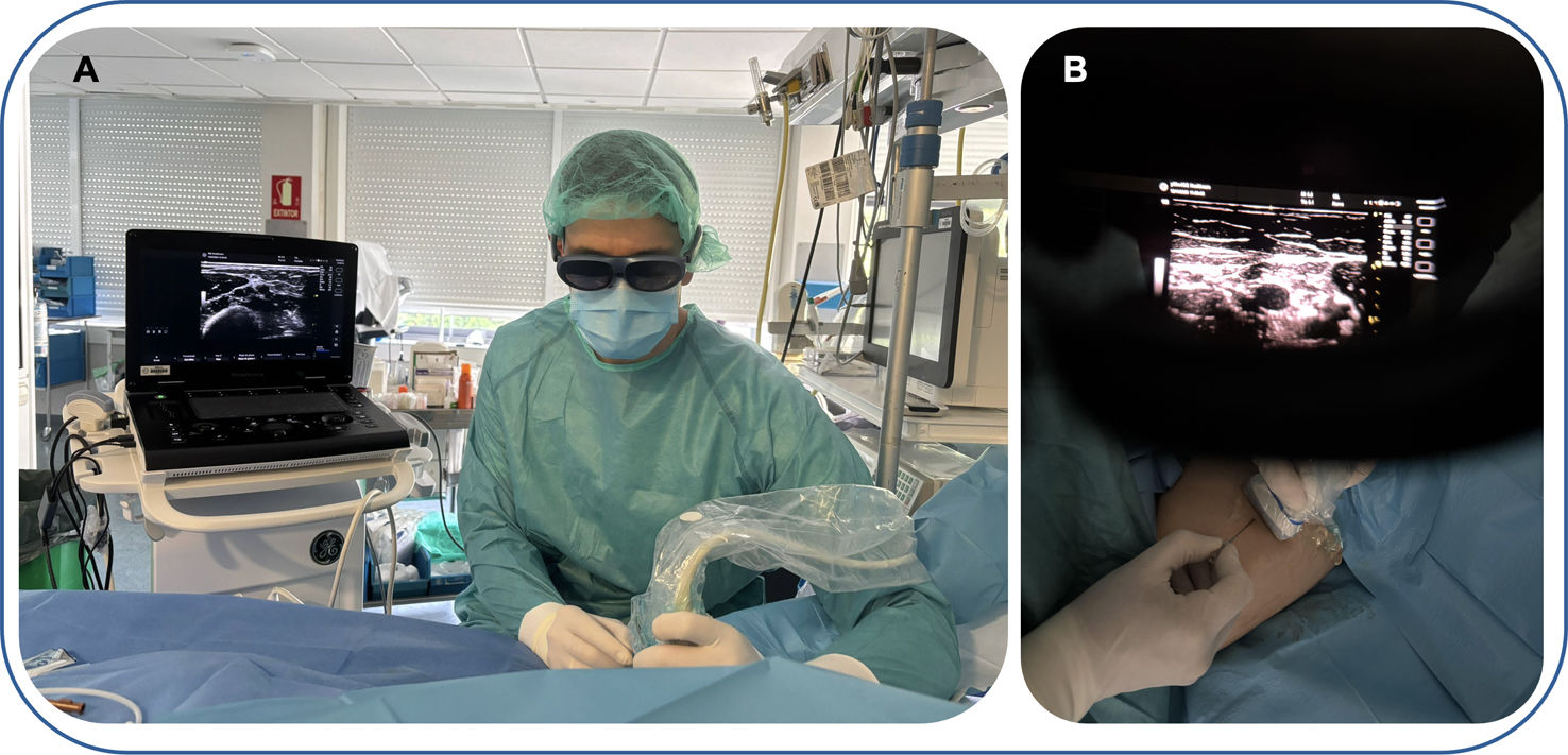
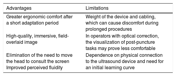
Our preliminary results indicate that image quality was satisfactory, with the main difference with respect to the conventional technique being the possibility of keeping a constant view of the puncture area, since the ultrasound image is projected in the upper part of the visual field without the need to turn the head (Fig. 1). This configuration offered distinct advantages, including enhanced concentration, improved coordination and greater flexibility in positioning the ultrasound machine within the cubicle. Table 1 shows the potential benefits and constraints in accordance with the perspective of the ICU nurse who served as the operator, in line with the aspects of competence outlined in the literature.7

Ultrasound-guided vascular cannulation procedure with augmented reality glasses (smart glasses). (A) Subjective operator view through the smart glasses while cannulating the venous access. (B) The ultrasound image is projected in the upper part of the operator's field of vision, eliminating the need to turn the head. This allows for uninterrupted viewing of the puncture area.
Operator-reported advantages and limitations of using augmented reality glasses in vascular cannulation.
| Advantages | Limitations |
|---|---|
| Greater ergonomic comfort after a short adaptation period | Weight of the device and cabling, which can cause discomfort during prolonged procedures |
| High-quality, immersive, field-overlaid image | In operators with optical correction, the visualization of post-puncture tasks may prove less comfortable |
| Elimination of the need to move the head to consult the screen Improved perceived fluidity | Dependence on physical connection to the ultrasound device and need for an initial learning curve |
Our preliminary experience suggests that this technical option offers advantages in terms of ergonomics and hand-eye coordination. It is therefore recommended that this topic be explored systematically and compared with the conventional technique. This would allow us to establish items for assessment, enabling us to define (based on both quantitative and qualitative parameters) its potential for the training of professionals and its application in clinical practice.8
Financial supportNo specific funding was received for this work.
Statement on the use of artificial intelligenceThe authors have not used any generative AI tools and/or AI-assisted technologies for the writing of this paper.
Author contributionsEVT, MARV, ARN, and ERR participated in the design of the project. EVT and ERR wrote and prepared the manuscript. MARV, ERR, and ARN supervised and approved the final version of the manuscript.
Conflicts of interestThe authors declare that they have no conflicts of interest.