

Article information
Full Text
Bibliography
Download PDF
Statistics
Figures (1)

Tables (1)
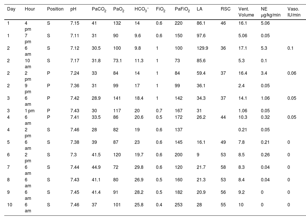
Table 1. Progression of internal environment, ventilatory and inotropic support.
Article
These are the options to access the full texts of the publication Medicina Intensiva (English Edition)
Member
If you are a member of the Sociedad Española de Medicina Intensiva, Crítica y Unidades Coronarias::
Go to the members area of the website of the SEMICYUC (www.semicyuc.org )and click the link to the magazine.
Go to the members area of the website of the SEMICYUC (www.semicyuc.org )and click the link to the magazine.

Purchase

