


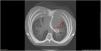
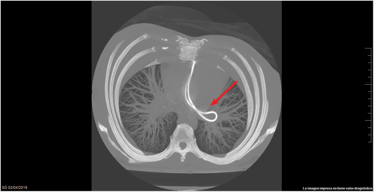
This is the case of a 36-year-old woman with myelomeningocele and hemorrhagic disease of the newborn who required ventriculoatrial drainage 11 years ago. She is admitted to the hospital with clinical signs of general discomfort, headache, and neck pain at the level of the scar of the ventriculoatrial valve (level of valve disconnection) of 5–6 clinical course. The CT scan performed reveals the significant dilatation of the supra and infratentorial ventricular system and sulci effacement (Fig. 1) with shunt valve with right frontal entry and tip on the left basal ganglia and interventricular shunt catheter and distal catheter migration towards the pulmonary artery (Fig. 2). Then, the catheter is extracted without complications at the cath lab using a snare. Afterwards, a new ventriculoatrial shunt is placed with good neurological outcomes.
Funding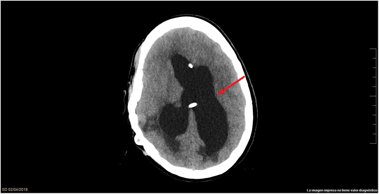
None whatsoever.
Please cite this article as: López Sánchez M, Domínguez Artiga MJ, Ortiz Lasa M. Migración catéter ventriculoatrial. Med Intensiva. 2022;46:296.

