


Our objectives were to describe the use of thromboprophylaxis and the incidence of VTE/bleeding in critically ill patients with hematologic malignancies (HM).
DesignRetrospective cohort study (2014–2022).
SettingMedic-Surgical Intensive Care Unit (ICU) in a tertiary care academic center.
PatientsAdult patients admitted to ICU with a concomitant diagnosis of a hematological malignancy.
InterventionsNone.
Main variables of interestWe analyzed demographic data, use of thromboprophylaxis and secondary outcomes that included incidence of VTE (venous thromboembolism), bleeding, mortality, severity scores and organ support. We applied a multivariable logistic regression model to examine the risk of thrombosis in the ICU.
ResultsWe included 862 ICU admissions (813 unique patients). Thromboprophylaxis was given during 65% of admissions (LMWH 14%, UFH 8%, and SCDs 43%); in 21% it was contraindicated due to thrombocytopenia; 14% of cases lacked documentation on prophylaxis. There were 38 unique incident cases of VTE (27 DVT, 11 PE), constituting 4.4% of ICU episodes. Most of VTE cases happened in patients with various degrees of thrombocytopenia. In the multivariable analysis, SOFA score on the first ICU day was independently associated (OR 0.85, 95% CI 0.76−0.96) with the risk of VTE. Bleeding occurred in 7.2% (minor) and 14.4% (major) of episodes; most frequent sites being CNS, abdomen/GI and pulmonary.
ConclusionsIn this cohort of critically ill patients with HM, there was considerable variability in the utilization of DVT prophylaxis, with predominant use of SCDs. The incidence of VTE was 4.4% and major bleeding 14%.
Clinical Trial RegistrationNCT05396157. Venous Thromboembolism in Hematologic Malignancy and Hematopoietic Cell Transplant Patients: a Retrospective Study (https://clinicaltrials.gov/).
Describir el uso de tromboprofilaxis y la incidencia de enfermedad tromboembólica y/o sangrado en pacientes críticos con enfermedad oncohematológica.
DiseñoEstudio retrospectivo de cohorte (2014–2022).
ÁmbitoUnidad cuidados intensivos medico-quirúrgica de un hospital terciario académico en Toronto, Canadá.
PacientesAdultos con diagnóstico de enfermedad oncohematológica ingresados en la UCI.
IntervencionesNinguna.
Variables de interés principalesSe analizaron los datos demográficos, el tipo de tromboprofilaxis utilizada y el riesgo de sangrado junto con los factores relacionados con éstos. Se aplicó un análisis de regresión logística multivariante para evaluar factores de riesgo para trombosis en la UCI.
ResultadosSe incluyeron 862 episodios de ingreso en la UCI (813 pacientes únicos). Se utilizó tromboprofilaxis en el 65% de los casos (heparina de bajo peso molecular 14%, heparina no fraccionada 8% y medios mecánicos 43%); en el 21% estuvo contraindicado por trombocitopenia y en el 14% de los casos no había documentación sobre profilaxis. Hubo 38 casos incidentes de enfermedad trombo embólica venosa o ETEV (27 trombosis venosa profunda, 11 embolismo pulmonar), lo que constituye el 4,4% de los episodios en la UCI. La mayoría de los casos de ETEV ocurrieron en pacientes con diversos grados de trombocitopenia. La puntuación SOFA en el primer día de UCI se relacionó de forma independiente en el análisis multivariable (OR 0.85, 95% CI 0.76−0.96) con el riesgo de ETEV. Se registró sangrado en el 7,2% (menor) y en el 14,4% (mayor) de los episodios; los sitios más frecuentes fueron el SNC, abdomen/GI y pulmonar.
ConclusionesEn esta cohorte de pacientes críticos con enfermedad oncohematológica, hubo una variabilidad considerable en la utilización de la profilaxis de la TVP, con un uso predominante de métodos mecánicos. La incidencia de ETEV fue del 4,4% y la de hemorragia mayor del 14%.
Venous thromboembolism (VTE) is a frequent, preventable, and potentially fatal complication in critically ill patients.1–3 The presence of active cancer, such as hematologic malignancy (HM), further increases thromboembolic risk.4,5 Patients with HM are at risk for both thrombosis and bleeding, which may be further increased in the setting of critical illness.4–6
Many patients with hematologic malignancy require admission to the Intensive Care Unit (ICU). Ferreyro et al. observed a 13.9% 1-year incidence of ICU admission for this population, with a median time from diagnosis to ICU admission of 35 days.7 In addition, Carini et al. recently highlighted a higher incidence of venous thromboembolism in critically ill patients with hematologic malignancy compared with those not requiring an ICU admission (3.7% vs 1.2% respectively).8 Clinical Practice Guidelines recommend pharmacological VTE prophylaxis in critically ill patients except in cases of high bleeding risk, when they recommend mechanical prophylaxis.9–13 Severe thrombocytopenia (platelets<50×109/L) is a frequent complication both in patients with HM and in critically ill patients, and is associated with worse outcomes and complications (i.e., higher bleeding risk).14–16 Regarding mechanical prophylaxis, it is not recommended as monotherapy except when pharmacological methods are contraindicated (grade 2A).9 Regarding bleeding, the same study by Carini et al. reported that 3.8% of included patients had at least one episode of major bleeding in the cohort, with a higher incidence in ICU patients compared to non-ICU hospitalized controls (7.6% vs 2.4%, OR 3.33; 95% CI 3.09–3.58).8 The balance between preventing VTE and minimizing bleeding complications underscores the complexity of managing thromboprophylaxis in critically ill patients with HM.
Given the increasing number of patients with hematologic malignancy who develop critical illness, it is imperative to understand the incidence and risk factors for venous thromboembolism, as well as the efficacy and risks associated with both chemical and mechanical thromboprophylaxis. Our objectives are to describe the: 1) use, type (mechanical or pharmacological) and timing of thromboprophylaxis; 2) incidence of catheter and non-catheter related VTE (deep venous thrombosis and pulmonary embolism) according to type of thromboprophylaxis (pharmacological or mechanical); and 3) incidence of bleeding episodes according to type of thromboprophylaxis (pharmacological or mechanical). We also described outcomes related to critical illness including ICU/hospital mortality.
Study design and methodsThe study was approved by the Research Ethics Boards at Mount Sinai Hospital (MSH) (#21-0109-C) and the University Health Network (#21-5476), who waived the need for consent. The study is registered with ClinicalTrials.gov (NCT05396157).
We conducted a single-center retrospective cohort study of adult patients (≥18 years) with a diagnosis of HM admitted to the ICU at MSH between January 1st, 2014, and January 31st, 2022. MSH is a tertiary care center which provides critical care services to Princess Margaret Cancer Center, the largest cancer center in Canada. There were no exclusion criteria. Each patient's relevant data was retrieved from the Electronic Health Record (EHR). Each ICU admission between hospital admission and discharge was included as a separate episode. Patients were followed until hospital discharge or death, whichever occurred first.
We report study findings in accordance with the STrengthening the Reporting of OBservational studies in Epidemiology (STROBE) checklist (eTable 1).17
Classification of hematologic malignancyThe type of HM was classified into categories: acute lymphoblastic leukemia (ALL), acute myeloid leukemia (AML), acute promyelocytic leukemia (APML), chronic lymphocytic leukemia (CLL), Hodgkin lymphoma and non-Hodgkin lymphoma, multiple myeloma, myelodysplastic syndrome, and others, following previously published studies.7,8
Outcomes and variablesFrom the EHR we obtained baseline characteristics including age, sex, previous VTE, comorbidities and type of HM. We recorded the lowest platelet value during the timeframe between ICU admission and ICU discharge. We defined categories of thrombocytopenia: moderate (50−99×109/L); severe (<50×109/L) and critical thrombocytopenia (<20×109/L).15,18 We calculated relevant severity scores (i.e., APACHE-II and SOFA) and recorded ECOG scores when available (eTable 2).19,20
VTE prophylaxisThe primary outcome was frequency of use, type (mechanical or pharmacological) and timing of thromboprophylaxis. At our institutions, pharmacological thromboprophylaxis is started as soon as possible for all inpatients, particularly patients admitted to the ICU, except for those with absolute contraindications including platelet counts<50×109/L, where mechanical methods are recommended.
We recorded the type of DVT prophylaxis during ICU admission and defined the following categories: pharmacological prophylaxis (either LMWH or UFH), mechanical (SCDs), and no prophylaxis, with an explicit contraindication documented in the EHR. If it was not possible to establish which type of prophylaxis the patient was receiving, or the reason prophylaxis was withheld, they were classified as “not stated”.
Thrombosis, bleeding and ICU related outcomesSecondary outcomes included the incidence of VTE during ICU admission (up to hospital discharge) and bleeding. We detected cases of DVT/VTE from the notes or from imaging results available in the EHR.21 The diagnostic criteria for DVT following published guidelines included the objective evidence of an intravascular clot, which could be visualized through duplex ultrasound imaging, contrast computed tomography (CT) venography, or contrast venography. Catheter-associated thrombosis was characterized as thrombosis occurring in the vein(s) where the catheter was positioned or in a nearby vein. Pulmonary embolism diagnosis was confirmed either by pulmonary angiography or contrast CT.21
We recorded all bleeding episodes and classified them according to the proposed International Society on Thrombosis and Haemostasis (ISTH) subtypes of minor or major bleeding.22 Major bleeding includes fatal bleeding, symptomatic bleeding in a critical area or organ, and bleeding resulting in a drop in hemoglobin level of ≥20g/L or leading to transfusion of ≥2 units of red blood cells.
Finally, we recorded variables independently associated with DVT or PE (i.e., history of VTE, type of HM, BMI, etc.)., together with relevant ICU outcomes including frequency and type of mechanical ventilation, vasopressor requirement, use of dialysis, and ICU and hospital mortality.23 We completed follow-up until hospital discharge, death, or transfer to another facility, whichever occurred first. We fitted a logistic regression model with the occurrence of thrombosis (DVT or PE) as the response variable, and various patient demographic and clinical characteristics as explanatory variables to describe potential risk factors for VTE.
Statistical analysisWe summarized baseline characteristics using median and interquartile range [IQR] for numeric variables, while for categorical variables we reported the percentage and associated raw counts. The incidence of VTE and major bleeding were described using proportions. Furthermore, we described the incidence of VTE related to platelet count and to the type of VTE prophylaxis the patient received in the ICU.
We provide numerical summaries of the main variables for all data, stratified by prophylaxis type and platelet count. Missing data was removed from calculations whenever information was unavailable. For numeric variables, we employed the Kruskal-Wallis non-parametric one-way ANOVA test, and for categorical variables, we employed the Pearson chi-square test of independence (with simulated exact critical values in cases of low cell counts).
We examined the simultaneous influence of multiple variables on the risk of thrombosis in the ICU. We fitted a logistic regression model with the occurrence of thrombosis (DVT or PE) as the response variable, and various patient demographic and clinical characteristics as explanatory variables. To identify a parsimonious model with good predictive behavior, we performed stepwise variable selection by optimizing Akaike’s Information Criterion (AIC).24 We included sex, age, BMI, and ICU length of stay as control variables, and 21 additional variables (eTable 3) that are described in the literature as risk factors for VTE to include in the model.25 Records with missing values over the considered variables were omitted, resulting in 754 complete data points for the control variables and the 21 potential predictor variables. The estimated coefficients of the selected model were transformed to equivalent Odds Ratios and plotted together with their associated confidence intervals and p-values.
All analyses were performed using the R Statistical Software version 4.3.1 (R Core Team 2023).26
ResultsBaseline characteristicsBetween January 1st, 2014, and January 31st, 2022, there were 7804 admissions in the ICU (see eFigure 1). Filtering based on admission diagnosis and premorbid conditions we identified 1022 ICU admissions of patients with concomitant hematologic malignancies (HMs), totaling 862 unique hospital admissions among 813 unique patients meeting inclusion criteria (see eFigure 2).
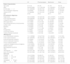
The median age was 61.5 years (IQR 50–69), with 58.8% males and a median BMI of 26 (IQR 24–30) (Table 1). The most frequent HMs were AML, ALL and lymphoma. About half of cases were diagnosed within a year prior to ICU admission, with 35% receiving hematopoietic cell transplantation (HCT) before ICU admission (81% within the preceding year). Thrombosis risk factors prevalence was 12.3% for a history of smoking, 10% for a history of VTE, 9.6% for concurrent cancer diagnosis, and 22.3% for a history of surgery before ICU admission. One third of patients had an ECOG status≥2.
Patient characteristics at baseline and according to thromboprophylaxis disease.
| All | Pharmacological | Mechanical | None | |
|---|---|---|---|---|
| Patient characteristics | ||||
| Age, years | 61.5 (19) | 63 (18) | 61 (18) | 59 (19) |
| Male | 58.8 (507/862) | 51.5 (100/194) | 61.3 (225/367) | 57 (103/180) |
| ECOG status≥2 | 30.1 (239/862) | 40.6 (67/194) | 26.5 (93/367) | 25 (40/180) |
| Non hematologic malignancy | 9.6 (83/862) | 10.3 (20/194) | 8.2 (30/367) | 11 (20/180) |
| History of VTE | 10.0 (86/856) | 21.5 (41/191) | 7.6 (28/366) | 6 (10/178) |
| BMI | 26 (6) | 26 (9) | 26 (7) | 26 (7) |
| ICU admission diagnoses | ||||
| Septic shock | 36.6 (316/862) | 33.5 (65/194) | 40.9 (150/367) | 31 (55/180) |
| Acute respiratory failure | 30.8 (266/862) | 33.5 (65/194) | 28.6 (105/367) | 38 (69/180) |
| Congestive heart failure | 2.6 (23/862) | 3.6 (7/194) | 2.7 (10/367) | 2 (4/180) |
| Multi-organ failure | 8.5 (74/862) | 6.2 (12/194) | 9.8 (36/367) | 3 (6/180) |
| Seizures/reduced level of consciousness | 6.2 (54/862) | 6.7 (13/194) | 5.5 (20/367) | 7 (12/180) |
| Cardiac arrest | 2.3 (20/862) | 2.6 (5/194) | 1.6 (6/367) | 2 (4/180) |
| Bleeding | 7.3 (63/862) | 2.1 (4/194) | 7.9 (29/367) | 13 (23/180) |
| Other | 3.1 (27/862) | 5.7 (11/194) | 1.6 (6/367) | 3 (6/180) |
| Postoperative | 0.7 (6/862) | 2.6 (5/194) | 0.2 (1/367) | 0 (0/180) |
| Pulmonary Embolus | 0.3 (3/862) | 1.0 (2/194) | 0 (0/367) | 1 (1/180) |
| Cytokine Release Syndrome | 1.1 (10/862) | 2.6 (5/194) | 1.09 (4/367) | 0 (0/180) |
| HM diagnosis | ||||
| Acute lymphoblastic leukemia | 15.1 (130/862) | 13.9 (27/194) | 13.1 (48/367) | 17 (31/180) |
| Acute myeloid leukemia | 48.7 (420/862) | 32.5 (63/194) | 54.5 (200/367) | 57 (103/180) |
| Acute promyelocytic leukemia | 3.5 (30/862) | 3.1% (6/194) | 2.7 (10/367) | 4 (8/180) |
| Chronic lymphocytic leukemia | 0.8 (7/862) | 2.1% (4/194) | 0.5% (2/367) | 0 (0/180) |
| Chronic myeloid leukemia | 3.0 (26/862) | 3.1% (6/194) | 3.5% (13/367) | 3 (5/180) |
| Lymphoma (Hodgkins and non-Hodgkins) | 14.4(124/862) | 29.9 (58/194) | 13.6 (50/367) | 4 (8/180) |
| Multiple myeloma | 5.5% (47/862) | 10.3 (20/194) | 3.5 (13/367) | 6 (10/180) |
| Myelodysplastic syndrome | 4.9 (42/862) | 3.6 (7/194) | 4.6 (17/367) | 3 (6/180) |
| Other | 4.2 (36/862) | 1.5 (3/194) | 3.8 (14/367) | 5 (9/180) |
| HM diagnosis<1 year | 45.5 (341/750) | 48.4 (76/157) | 44.9 (153/341) | 42 (55/131) |
| Stem cell transplant | 35.0 (301/860) | 31.4 (61/194) | 39.6 (145/367) | 28 (50/180) |
| Cell count | ||||
| Platelets≥50 | 19.4 (167/862) | 59 (98/167) | 15 (25/167) | 26 (44/167) |
| Platelets≥20 & <50 | 23.1 (199/862) | 26.8 (52/194) | 19.9 (73/367) | 24 (44/180) |
| Platelets<20 | 57.5 (496/862) | 22.7(44/194) | 73.3 (269/367) | 62 (112/180) |
| Hemoglobin<70 (g/L) | 53.0 (457/862) | 31.4 (61/194) | 60.8 (223/367) | 57 (102/180) |
| WBC<4×10⁹/L | 52.3 (451/862) | 29.9 (58/194) | 61.3 (225/367) | 56 (100/180) |
Data are presented as median (IQR) or % (n/N) conditional on the column category.
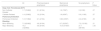
DVT prophylaxis included pharmacological strategies in 22.6% of episodes, mechanical strategies in 42.6% and in 34.9%, prophylaxis was not used or mentioned. Among patients without documented prophylaxis, 60.8% had critical thrombocytopenia (Table 2).
DVT prophylaxis according to platelets count.
| DVT prophylaxis | All ICU admissions | Platelets<50 | Platelets<20 |
|---|---|---|---|
| None | 180 (20.9) | 44 (22) | 112 (23) |
| Low Molecular Weight Heparin | 119 (13.8) | 32 (16) | 27 (5) |
| Unfractioned heparin | 71 (8.2) | 19 (10) | 17 (3) |
| Oral anticoagulants | 4 (0.5) | 1 (0.5) | 0 (0) |
| Sequential compression devices | 367 (42.6) | 73 (37) | 269 (54) |
| Compression stocking | 0 (0) | 0 (0) | 0 (0) |
| Not Stated | 121 (14) | 30 (15) | 71 (14) |
| Total | 862 (100) | 199 (100) | 496 (1) |
Data are presented as N (%) conditional on the column category.
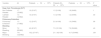
During or following ICU admission, there were 38 unique incident cases of VTE (27 DVT, 11 PE), constituting 4.4% of ICU episodes. Of the DVT cases, 15 were non-catheter related (80% in the lower extremities) and 12 were catheter-related (90% in the upper extremities). Notably, 29.6% and 48.1% of DVT cases were receiving pharmacological or mechanical prophylaxis, respectively (Table 3). Thrombosis risk factors included prior DVT (7.5%) and previous PE (3.7%). eFigure 3 shows the incidence of DVT and PE per year. Of note, 66.7% of patients with pre-ICU DVT and 61.3% of patients with PE had moderate thrombocytopenia at the time of ICU admission.
Thrombosis and Bleeding by VTE Prophylaxis.
| All | Pharmacological prophylaxis | Mechanical prophylaxis | No prophylaxis | P Value | |
|---|---|---|---|---|---|
| Deep Vein Thrombosis (DVT) | |||||
| Non-Catheter Related | 1.7 (15/862) | 3.1 (6/194) | 1.9 (7/367) | 0 (0/180) | .07 |
| Catheter Related | 1.4 (12/862) | 1.0 (2/194) | 1.6 (6/367) | 0.6 (1/180) | .57 |
| Pulmonary Embolism | |||||
| Pulmonary Embolism | 1.3 (11/862) | 2.1 (4/194) | 0.82 (3/367) | 2.2 (4/180) | .33 |
| Bleeding | |||||
| Minor Bleeding | 7.2 (62/862) | 4.6 (9/194) | 8.7 (32/367) | 7.2 (13/180) | .21 |
| Major Bleeding | 14.2 (122/862) | 4.6 (9/194) | 15.5 (57/367) | 21.1 (38/180) | <.05 |
Data are presented as % (N/total) conditional on the column category.
Bleeding episodes were classified them according to the proposed International Society on Thrombosis and Haemostasis (ISTH) subtypes of minor or major bleeding.22 Major bleeding includes fatal bleeding, symptomatic bleeding in a critical area or organ, and bleeding resulting in a drop in hemoglobin level of 2g/L or leading to transfusion of 2 or more units of red blood cells, minor bleeding being any other type of bleeding.
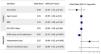
We used a logistic regression model to examine the combined effects of multiple variables on the risk of VTE. We included patient sex, age, BMI, and ICU length of stay as controls, and selected three additional variables that help predict the incidence of VTE (according to Akaike’s Information Criterion; AIC).27 The resulting odds ratios with associated confidence intervals are presented in Fig. 1. In the multivariable analysis, the only variable that remained significantly associated with VTE was the SOFA score (P=.007) on the first ICU Day, which lowers the risk of VTE by 15% per each unit of the scale, implying patients in more severe condition were less likely to be diagnosed with VTE. Vasopressors and plasma transfusions are the other two selected variables, with no significant association in our series (P=.06 and P=.078 respectively).

Bleeding occurred in 7.2% (minor) and 14.4% (major) of episodes. Table 3 shows the incidence of VTE and bleeding outcomes across this population by type of DVT prophylaxis. The most frequent bleeding sites for major bleeding were central nervous system (CNS) (44.3%), abdomen/gastrointestinal (GI) (29.5%), and pulmonary (19.7%).
Thrombosis and bleeding related to thrombocytopeniaInterestingly, 60% of non-catheter related DVT, 50% of catheter related DVT and 45.4% of PE cases respectively, occurred in patients with critical thrombocytopenia. The median platelet count at DVT/PE diagnosis was 54 (IQR 22–133) (Table 4).
Thrombosis and Bleeding by Platelet counts.
| Variable | All | Platelets>50×109/L | Platelets 20–50×109/L | Platelets<20×109/L | P value |
|---|---|---|---|---|---|
| Deep Vein Thrombosis (DVT) | |||||
| Non-Catheter Related | 1.7 (15/862) | 1.8 (3/167) | 1.5 (3/199) | 1.8 (9/496) | 1 |
| Catheter Related | 1.4 (12/862) | 1.8 (3/167) | 1.5 (3/199) | 1.2 (6/496) | .93 |
| Pulmonary Embolism | |||||
| Pulmonary Embolism | 1.3 (11/862) | 1.2 (2/167) | 2.0 (4/199) | 1.0 (5/496) | .66 |
| Bleeding | |||||
| Minor Bleeding | 7.2 (62/862) | 4.8 (8/167) | 7.5 (15/199) | 7.9 (39/496) | .40 |
| Major Bleeding | 14.1 (122/862) | 16.2 (27/167) | 21.1 (42/199) | 10.7 (53/496) | .001 |
Data are presented as % (N/total) conditional on the column category.
DVT: deep venous thrombosis; VTE: Venous thromboembolism.
Regarding bleeding, 62.9% of minor bleeding cases and 43.4% of major bleeding cases occurred in patients with critical thrombocytopenia (Table 4). 60% patients required platelet transfusion (median 3 units, IQR 1–5), 68% packed red blood cell (PRBC) transfusion (median 2 units, IQR 1–5), and 14% fresh frozen plasma (FFP) (median 3 units, IQR 1–5).
ICU diagnosis, procedures, and mortalityThe main reasons for ICU admission were septic shock and acute respiratory failure. Regarding organ support, more than half of patients required invasive mechanical ventilation, 3.9% non-invasive ventilation (NIV), and 9% high-flow oxygen therapy (eTable 4). Additionally, more than half of patients required vasopressor support and 12.1% dialysis, and the median SOFA score was 8 (IQR 6–10). Median pre-ICU hospital length of stay (LOS) was 7 days (IQR 1–19), median ICU LOS was 3 days (IQR 1–6), and median total hospital LOS was 30 days (IQR 15–51).
Regarding thromboprophylaxis and ICU related variables, patients receiving mechanical prophylaxis had higher rates of invasive mechanical ventilation (62%, P=.0003), requirement for vasopressors (62%, P=.02), requirement for dialysis (17%, P=.002), higher SOFA and APACHE-II, and higher ICU mortality (35%, P=.03).
Overall ICU mortality was 34.1%, and hospital mortality in ICU survivors was 29.7%. ICU mortality was 25.9% for patients with DVT and 45.4% for patients with PE; hospital mortality was 45% for patients with DVT and 0% for patients with PE. ICU mortality was 40.2% and hospital mortality was 31.5% for patients with major bleeding.
DiscussionThis cohort study of 813 unique patients (accounting for 862 unique hospital admissions) represents the largest analysis of the incidence of and risk factors associated with VTE and bleeding among patients with HM admitted to ICU, and on thromboprophylaxis strategies in this population. Remarkably, we found that in more than one third of episodes prophylaxis was not used or not documented and that there was a non-negligible incidence of VTE and/or bleeding during ICU admission.
Effectively implementing DVT prophylaxis is challenging in some clinical settings. In a previous multinational cross-sectional survey only 37% of patients with active malignancy received adequate prophylaxis, with thrombocytopenia as one of the associated factors with inadequate prophylaxis.28 In the ICU, there is a reported 70% adherence to guideline recommendations, however there is also a lack of mechanical devices in cases where pharmacological prophylaxis is contraindicated as reported by García-Olivares.29,30
Thrombocytopenia does not appear to be strictly protective against VTE, and previous studies document the safety of prophylactic enoxaparin even in patients with platelets lower than 10×109/L.5,31 In addition, the omission of thromboprophylaxis within the first 24h of ICU admission without clinical reasons has been associated with increased mortality in critically ill adults.32 A notable finding in our study is the substantial proportion of patients not receiving any prophylaxis or in which there was no mention of it. There is extensive literature around inadequate VTE prophylaxis that helps to better frame this issue. In a previous cross-sectional study, the factors associated with an increased risk for inadequate VTE prophylaxis included the presence of a contraindication to pharmacological prophylaxis (OR 3.9, 95% CI 2.5–6.1), and ICU admission for a non-medical pathology (OR 11.1; 95% CI 7.6–16.1).33 In a retrospective analysis of 1.4 million critically ill patients, 4% did not receive thromboprophylaxis within the first 24h after ICU admission, without obvious reason.29 This is a lower proportion than our study, where thromboprophylaxis was withheld in 21% of admissions because of thrombocytopenia; and there was no mention of prophylaxis in 14%. In the ENDORSE study, platelet count<100×109/L was a contraindication for VTE prophylaxis.28 Furthermore, up to 50% of all hospitalized patients at risk for VTE were not receiving appropriate prophylaxis. The use of prophylaxis protocols has been proven to be feasible and could potentially reduce the risk of VTE in this population.34,35
Finally, we found a non-negligible incidence of bleeding (7% minor bleeding and 14% major bleeding). This rate is higher than the reported in a previous population study that found a higher incidence of bleeding in ICU patients compared to non-ICU hospitalized controls (7.6% vs 2.4%, OR 3.33; 95% CI 3.09–3.58).8 In an analysis of factors associated with bleeding risk in medical patients, Decousus et al. reported a cumulative 3.2% incidence of major and nonmajor in-hospital bleeding within 14 days of admission. Active gastroduodenal ulcer (OR, 4.15; 95% CI, 2.21–7.77), prior bleeding (OR, 3.64; 95% CI, 2.21–5.99), and low platelet count (<50×109/L) (OR, 3.37; 95% CI, 1.84–6.18) were the strongest independent risk factors at admission for bleeding.36
As a retrospective analysis, this study has several limitations. Detection of events was done by individual chart review, subjected to human error. Also, there was lack of documentation for prophylaxis in 14% of cases which could potentially influence the results, either underestimating or overestimating the thromboprophylaxis use. Furthermore, despite being conducted at Canada’s largest HM centre, this was a single-center study. Further, VTE events are limited to those where the diagnosis was clinically suspected and a confirmatory test done, possibly leading to under-detection compared with active surveillance ultrasound for DVT.37 Finally, we lack data on some risk factors for VTE, such as previous chemotherapy. The strengths of our study include the large HM population, and the detailed and comprehensive data collection, including information regarding ICU-related treatments (i.e., mechanical ventilation, vasopressor requirements, etc.) and severity scores. By examining individual patient records instead of solely relying on administrative data, we could determine details about VTE timing and platelet counts.
We anticipated a low VTE risk in patients with HM and thrombocytopenia, however we found a considerable rate of thrombosis considering that this was a non-surveillance scenario. The risk of thrombosis was similar to that observed in a non-HM population of critically ill patients receiving chemical thromboprophylaxis.38 Additionally, our study revealed a high risk of bleeding, surpassing rates observed in general ICU populations, where studies report rates of major bleeding closer to 5%.39
ConclusionsThe risk of VTE in critically ill patients with HM should be considered even in patients with thrombocytopenia. The risk of bleeding was considerably higher than expected. Our results should inform future prospective studies on this topic to try and better define the risk of thrombosis and bleeding in this population in a surveillance context.
Conflict of interestThe authors have no relevant financial or non-financial interests to disclose. The authors have no conflicts of interest to declare that are relevant to the content of this article.
Funding informationFCC was awarded the Hold’em for Life Oncology Fellowship 2021-2022 for the completion of this project.
Notation of prior abstract publication- a
Poster presentation at Critical Care Canada Forum 2022 (Carini FC, Dozois G, Novitzky-Basso I, Munshi L, Ferreyro B, Mehta S. Critical Care Canada Forum 2022 Abstracts. Canadian Journal of Anesthesia/Journal canadien d’anesthésie. 2023;70(1):s175-s177. doi:https://doi.org/10.1007/s12630-023-02445-y).
- b
Presented as a poster at the Virtual Art Slutsky Research Day, Interdepartmental Division of Critical Care Medicine, University of Toronto, June 21, 2022.
The study was approved by the Research Ethics Boards at Mount Sinai Hospital (#21-0109-C) and the University Health Network (#21-5476). We requested a full waiver of consent and Personal Information Protection and Electronic Document Act (PIPEDA) authorization for the patients identified in this study.
Statement of generative AI and AI-assisted technologies in the writing processDuring the preparation of this work, the author used ChatGPT, an AI language model developed by OpenAI, in order to improve readability and to check grammar and spelling. After using this tool, the author reviewed and edited the text and assume full responsibility for the content of the publication.
FCC had full access to the data and takes responsibility for the integrity of the data and accuracy of the data analysis. Concept and design: FCC, BLF, SM, INB and LM. Acquisition, analysis, and interpretation of data: FCC, SM, SD. Drafting of the manuscript: FCC, CH and SM. Critical revision of the manuscript for important intellectual content and approval of the final draft: all authors. Statistical analysis: SD, FCC.
We thank Mr. Anaqi As Shafiq Bin Amir Razif (anaqi.amirrazif@mail.utoronto.ca, University of Toronto Scarborough) for his collaboration in the statistical analysis for this project.
The following is Supplementary data to this article:


