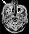
A 72-year-old male presented with a history of advanced type 2 diabetes and the fitting of an upper dental bridge four months ago. He was admitted due to pain, facial swelling, palpebral ptosis, unstable gait and septic shock. The facial/sinuses CAT scan revealed right-side ethmoid-maxillary sinus disease. The axial MRI study showed right maxillary sinus occupation with a hypointense central zone in T2-weighted sequencing (*, Fig. 1), consistent with fungal sinusitis/mucormycosis extending to the orbital region (→, Fig. 1). Focal restricted diffusion was observed in the right frontal zone, showing enhanced signal intensity in diffusion (b1000) (Fig. 2a) and a hypointense signal in ADC mapping (Fig. 2b), in relation to cerebritis. Based on these findings, antimicrobial and antifungal treatment was provided, and extensive surgical resection of the accessible lesions was performed. The pathology report confirmed the diagnosis of mucormycosis. The clinical course proved favorable, and the patient was discharged after 33 days of hospital stay.
Conflicts of interestThe authors have no conflicts of interest to declare.
Thanks are due to all the staff involved in the care of the patient, and to all those who made the drafting and publication of this article possible.
Please cite this article as: Vallverdú Vidal M, Iglesias Moles S, Palomar Martínez M. Mucormicosis rino-órbito-cerebral en un paciente crítico. Med Intensiva. 2017;41:509–510.


