Información de la revista
Vol. 47. Núm. 9.
Páginas 556-557 (Septiembre 2023)
Images in Intensive Medicine
Acceso a texto completo
Esophageal prosthesis migrated to gastric corpus. An ultrasound diagnosis
Prótesis esofágica migrada a cuerpo gástrico. Un diagnóstico ecográfico
Visitas
2038
Adela Fernández Galileaa,
Autor para correspondencia
adelafer_90@hotmail.com

Corresponding author.
, Juan Francisco Muñoz Morenob, Raúl Vicho Pereirac
a Servicio de Medicina Intensiva, Hospital General Virgen Macarena, Sevilla, Spain
b Servicio de Medicina Intensiva, Hospital General Universitario de Ciudad Real, Ciudad Real, Spain
c Servicio de Medicina Intensiva, Clínica Rotger Quirónsalud-Hospital Mallorca, Palma, Balearic Islands, Spain
Este artículo ha recibido
Información del artículo
Texto completo
Descargar PDF
Estadísticas
Figuras (3)
Mostrar másMostrar menos
Texto completo

This is the case of a patient admitted to the intensive care unit (ICU) due to early suture dehiscence during the postoperative period of a surgical myotomy performed due to an esophageal diverticulum. A metal self-expanding prosthesis is placed endoscopically in the dehiscence region.

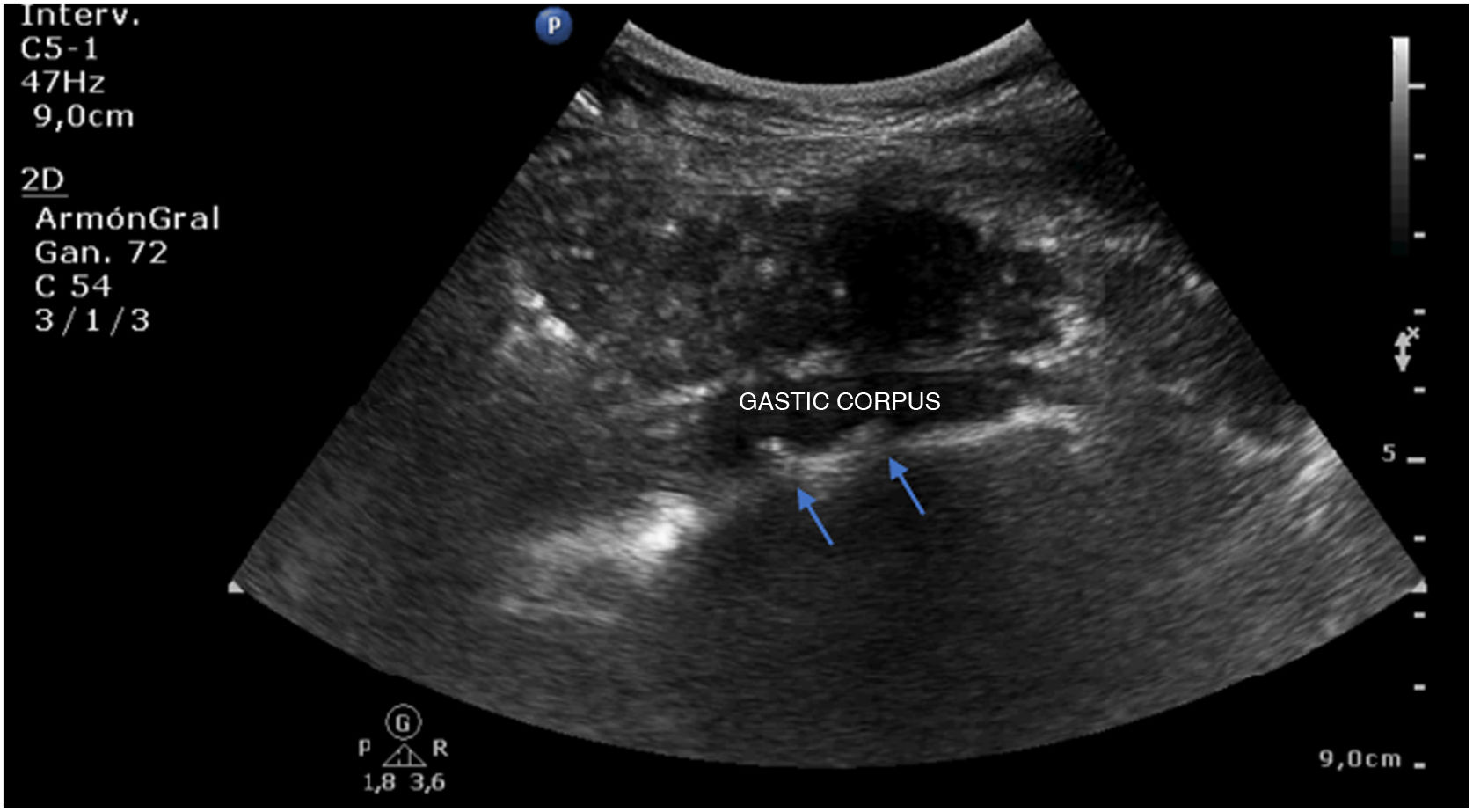
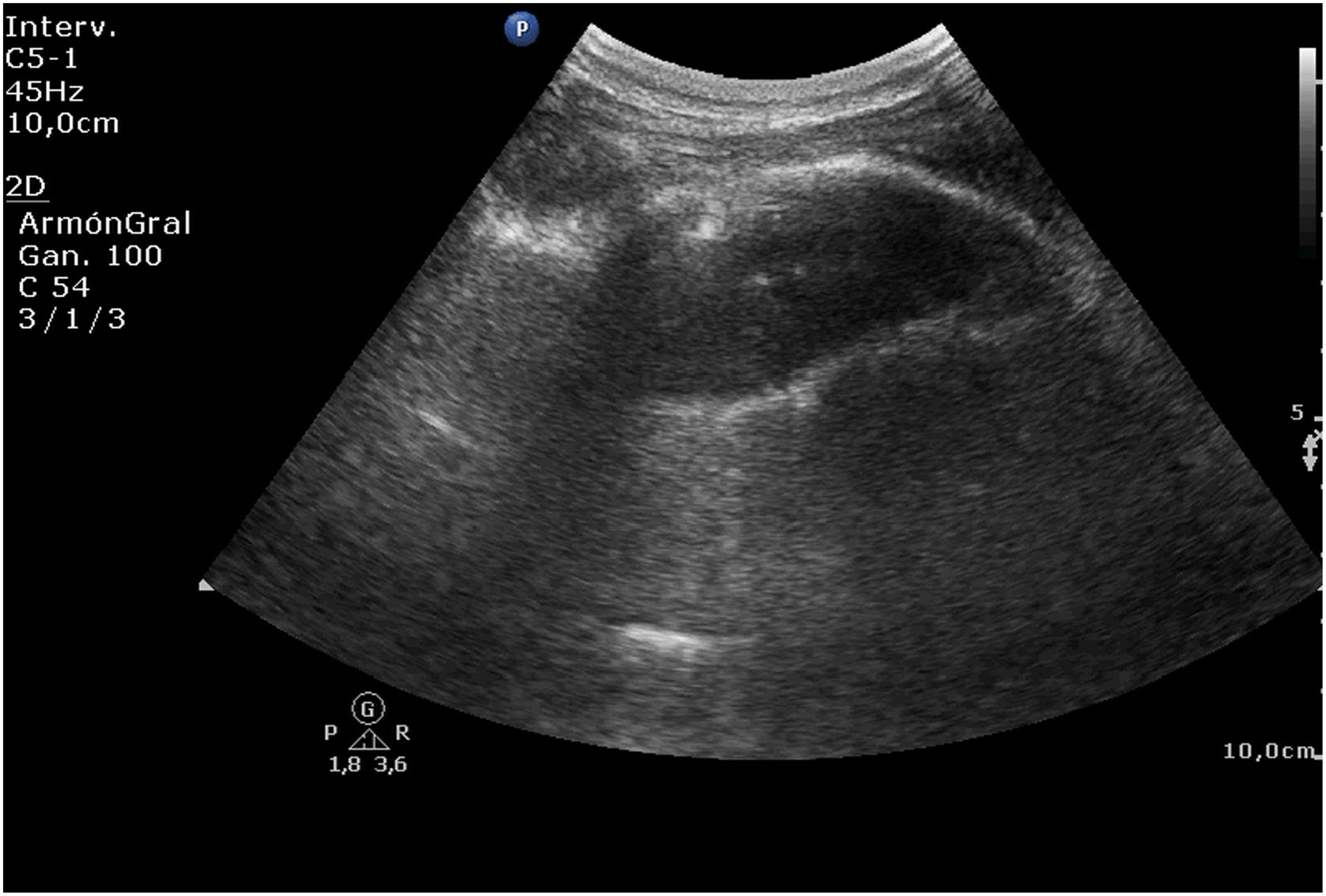
The ultrasound follow-up conducted a few hours later shows the endoprosthesis (Fig. 1) with its “clips” (arrows in Fig. 2) at gastric corpus region in the splenorenal window. Through the endoscopy the prosthesis is repositioned until it reaches the proper location. The ultrasound follow-up confirms the disappearance of the device in the stomach (Fig. 3).

Figure 1
Figure 2
Figure 3
Authors’ contributions

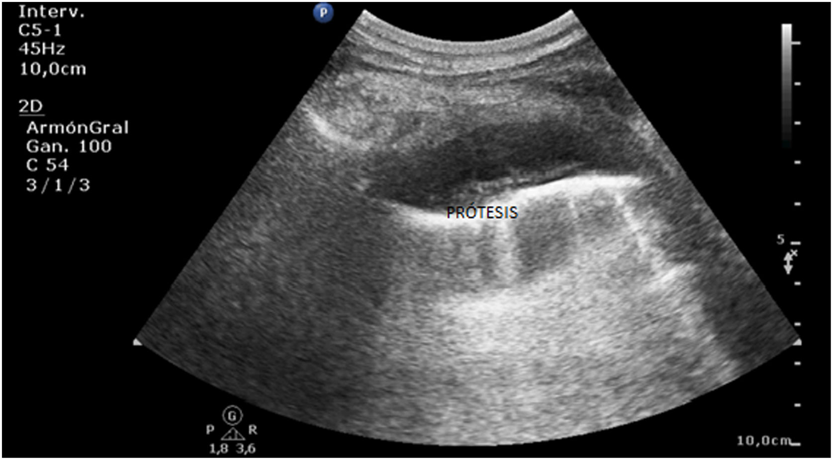
All the authors were similarly involved in the diagnosis and management of the patient, and the design and drafting of this manuscript.

Copyright © 2023. Elsevier España, S.L.U. and SEMICYUC
Descargar PDF
Idiomas
Medicina Intensiva
Opciones de artículo
Herramientas