

Cerebral microdialysis, introduced in experimental studies 40 years ago, has been used clinically since 1992 for the neurochemical monitoring of patients in intensive care. The principles underlying this technique are closely related to brain metabolism. The study of the metabolites detected at brain interstitial tissue level, through the semipermeable membrane of the device, allows us to assess different physiological pathways in the brain, analyzing the changes that occur when they become less efficient in terms of energy, and also detecting waste products secondary to tissue damage. Despite its current limitations, this technique provides relevant information for research and the clinical management of critical neurological patients.
La microdiálisis cerebral, introducida en estudios experimentales hace unos 40 años, ha sido empleada en la clínica desde 1992 para la monitorización neuroquímica de pacientes en unidades de cuidados intensivos. Los principios en los que se basa esta técnica se encuentran íntimamente ligados al metabolismo cerebral. El estudio de los metabolitos detectados en el intersticio tisular cerebral, a través de la membrana semipermeable de la que dispone el dispositivo, permite estimar la situación de las distintas rutas metabólicas fisiológicas cerebrales, analizando las modificaciones que se producen cuando estas tornan hacia rutas menos eficientes desde el punto de vista energético y detectando productos de desecho secundarios a la lesión tisular. A pesar de sus limitaciones actuales, esta técnica aporta información relevante para la investigación y el abordaje clínico de los pacientes neurocríticos.
Cerebral microdialysis (CMD), one of the latest neuromonitorization techniques introduced in the study of neurocritical patients, is of great clinical interest and can be used at the patient bedside. The analysis of the results of CMD informs us of the brain metabolic conditions in the context of serious neurological disorders, affording valuable information in certain neurocritical situations.
Cerebral hypoxia produced by different disorders of cerebral or systemic origin is a common situation, and is one of the leading causes of secondary damage in patients with acute brain conditions. In the course of the scientific and technological developments in neurocritical care units, a number of variables have been used to ensure the early detection of cerebral hypoxia, with the purpose of avoiding or preventing the problem, or—in extreme cases—of palliating its consequences. Techniques such as the measurement of intracranial pressure (ICP), cerebral perfusion pressure (CPP), cerebral blood flow (CBF), regional oxygen saturation (using near-infrared spectroscopy (NIRS)) and cerebral tissue oxygen pressure (PtiO2) are all widely used in the neurointensive care setting.1–3
These techniques offer information on the regional or global hemodynamic situation of the brain and on oxygen availability in the brain tissue, and this in turn conditions our approach in relation to the healthy and damaged parenchyma.4,5 However, the intensity and repercussions of these altered variables upon cell metabolism, and adjustment to the existing oxygen deficit in each individual patient, remain unclear. It is known that a cascade of biochemical reactions, resulting from the lack of balance between energy demand and supply,6 gives rise to conversion from aerobic cell metabolism to anaerobic metabolism, where ATP is produced less efficiently, and where cell destruction is the norm. Through a number of physiopathological mechanisms, the perpetuation of this metabolic change results in what we now know as secondary brain damage. It is at this point where CMD can prove useful. Thanks to this technique, we can obtain information at the patient bedside referred to cell metabolism and end cellular degradation7 in situations of brain damage, based on the use of energy substrates and on the production of different metabolites and neurotransmitters.
The present study offers a brief review of this technique and its contribution to the management of neurocritical patients.
Historical reviewAlthough the concept of dialysis is by no means new, its first application to brain studies in animals was made by Bito et al.8 around 1966. These studies gave rise to the idea that the cerebral extracellular compartment maintains a composition different from that of both cerebrospinal fluid (CSF) and plasma. Posteriorly, in 1972, Delgado and DeFeudis,9 likewise in laboratory animals, developed the concept of continuous microdialysis (MD). However, it was not until 1974, following the work of Ungerstedt and Pycock,10 when the model conceived by Delgado and DeFeudis9 was improved with a view to obtaining reliable and analyzable cerebral microdialysate samples. This important step in turn supported the posterior investigations of Ungerstedt,11 who began to use CMD in humans around 1982. The search for a biochemical pattern reflecting healthy and diseased brain function explains the subsequent explosion of scientific events in the field of CMD. The use of this technique, initially conceived as a laboratory aid, has allowed CMD to play a relevant clinical role since 1992, when it was introduced as a neurochemical monitorization tool at the bedside of patients admitted to the neurocritical care unit.12
Principles of cerebral microdialysisThe principles on which this technique is based are intimately related to brain metabolism. The study of the metabolites detected in the tissue interstice through the semipermeable membrane of the device, allows us to assess the conditions of the different cerebral physiological pathways, analyzing the changes that occur when these pathways shift towards energetically less efficient mechanisms, and to detect waste products secondary to tissue damage.13,14
More specifically, MD allows us to monitor the following metabolites:
- -
Metabolites related to energetic and metabolic cascades: glucose, pyruvate and lactate.
- -
Excitatory amino acids: glutamate.
- -
Markers of cell membrane damage: glycerol.
- -
Markers of protein alterations: urea.
- -
Other substances: cytokines, nitric oxide (NO) metabolites, N-acetylaspartate (NAA), etc.
Microdialysis is based on the capacity of solutes to cross a semipermeable membrane until an equilibrium is reached on either side of the membrane. To this effect, use is made of a catheter consisting of a double-lumen probe coated at its tip by a dialyzing semipermeable membrane. The probe is implanted in the brain tissue, and an isotonic solution of known characteristics is then perfused through one of the two channels at a programmable rate (0.1–5μl/min). Once this solution comes into contact with the tissue interstice, substance exchange takes place through the semipermeable membrane, and the second catheter channel accumulates a liquid with a composition different from that of the original liquid, known as the microdialysate. The diffusion mechanisms allow the molecules to cross the membrane, based on the existing concentration gradient. In this way, those molecules that are sufficiently small to cross the membrane and which are present in high concentrations within the interstice, become part of the microdialysate, with a minimum passage of water. The concentration gradient is maintained thanks to the fact that the perfusion fluid flows constantly.15
The MD catheter collects the microdialysate in accordance to the characteristics of the cerebral extracellular compartment (tissue interstice). This simple concept offers a technique with many potential applications to any molecule sufficiently small to cross the membrane. Until recently, only substances such as glucose, urea, lactate, pyruvate and glycerol, with molecular weights of under 20kDa, could be studied, since the polyamide membrane employed could only be crossed by molecules below this limit.16 At present, however, we have membranes that can filter molecules of up to 100kDa. In this context, De los Rios et al.17 have published a study analyzing the usefulness of high-resolution CMD in recovering inflammatory mediators and proteins (IL-1β, IL-6, IL-8, IL-10, TNFα, MMP2 and MMP9) from the microdialysate.
In examining the behavior of a given solute, and assuming an adequate molecular size, the intracatheter quantities of the solute will depend on its concentration in the interstitial space, the diffusion coefficient of the substance, the properties and pore size of the membrane, and the catheter liquid perfusion rate involved. Based on these parameters, the percentage concentration of the intracatheter solute is calculated with respect to the actual extracellular concentration, in what is known as the recovery rate. This rate is dependent upon the mentioned stable parameters (pore size, membrane length and infusion rate) and also on other dynamic or unpredictable variables (temperature of the brain tissue, characteristics of interstitial diffusion, size of the interstitial compartment, and the presence of gliosis induced by insertion of the catheter). The importance of these dynamic variables is that they do not allow us to establish the recovery rate once the catheter has been inserted—changes in these variables being able to alter the recovery rate. However, the in vivo determination of extraneuronal urea has given us an approximation, since the brain and adipose tissues have the same concentration of urea, with an excellent correlation during 6 days between both dialysates—this allowing us to assume a small and stable (though individual) influence of the cerebral dynamic variables upon the brain recovery rate, particularly as regards substances with diffusion characteristics similar to urea such as glutamate, lactate and pyruvate. Monitorization of the ratio between the concentration of urea in the neuronal and subcutaneous interstices is also of a clear practical use, allowing us to assess correct microcatheter function. In the absence of changes in the mentioned recovery rate, we can guarantee that the variations in brain metabolites in the dialysate offer an artifact-free indication of the corresponding variations in the interstitial space. This indicates that it is advisable to place a second microdialysis catheter in the subcutaneous cellular tissue as a guide in assessing the changes in metabolites occurring at brain and systemic level.
In order to determine the recovery rate of a solute, for a given membrane and perfusion rate, we submerge a microcatheter in a standard solution of the study substance and determine the difference between its intra- and extracatheter concentration. Thus, for a microcatheter with a pore size of 20kDa and a length of 10mm, and assuming a perfusion rate of 2μl/min, the recovery rate is 34% for glutamate, 62% for lactate, and 63% for pyruvate. Lower perfusion rates and larger membrane surfaces result in higher recovery rates. Specifically, at a flow rate of 0.3μl/min and with a membrane length of 30mm (i.e., the CMD specifications usually found in clinical practice), recovery is close to 100%.
In the same way as with other local monitoring systems such as PtiO2 or NIRS, monitorization with the MD catheter encompasses a small area of the brain (approximately 1.5cm2), and this may represent a limitation of the technique. It is therefore advisable to implant the catheter in zones at risk of developing secondary damage (shadow zones, zones of possible vasospasm, around space-occupying lesions, etc.), since these areas will better reflect the pathological brain metabolism. In cases of diffuse axonal damage, it is advisable to position the catheter in the non-dominant hemisphere. Adequate catheter positioning is checked by locating its radiopaque gold tip by computed tomography.
Brain metabolismIn terms of energy metabolism, the brain represents only 2% of the total body weight, but is responsible for 20% of global oxygen consumption and 25% of total glucose consumption, and moreover receives 15–20% of the cardiac output under resting conditions. Fifty-five percent of this high energy consumption corresponds to the generation of electrical signals, and 45% to metabolic processes—including membrane stabilization, ion pump operation and the synthesis of structural and functional molecules. In addition, the fact that the brain shows no pauses in demand means that a practically constant metabolic rate must be maintained day and night. Furthermore, while other tissues are able to operate for short periods of time without oxygen supply, the brain is totally dependent upon oxidative metabolism.18
In this sense, and in contrast to other organs that can use different substances (lipids, amino acids and different sugars) as an energy source, the brain is nurtured exclusively by blood glucose. Recent studies have revealed a certain degree of compartmentalization in the brain energy processes, whereby in the event of a point increase in neuron activation, potassium, glutamate and other neurotransmitters are released within the extracellular space—generating an increase in astrocyte activity destined to restore the composition of the cerebral cortical microenvironment. In order for this to occur, astrocyte hyperglycolysis is required, giving rise to an increase in the extracellular concentrations of lactate, which in turn is taken up by the neurons to close this metabolic circle.
From all these processes it is deduced that the brain is completely dependent upon the bloodstream to secure a continuous supply of oxygen and glucose, and that cerebral blood flow is closely related to local metabolic expenditure—increasing or decreasing according to the local brain demands. This regulatory mechanism may be interrupted by lesions, causing the brain to become more vulnerable to secondary ischemic-metabolic damage. However, there are aspects of brain metabolism that have not been fully clarified. In this sense, alterations have been observed in the values of the metabolites in the microdialysate similar to those found in brain ischemia, with both CBF and PtiO2 remaining within normal ranges. As a possible explanation, it has been postulated that other metabolic pathways and associated enzymes may be involved, independent of oxygen and glucose supply to the cell.19 Further research in this field therefore appears necessary.
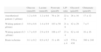
Metabolites commonly studied by cerebral microdialysisAs has been commented, CMD is able to analyze a broad range of metabolites and mediators of brain damage. However, the greatest clinical experience has been gained with those listed in Table 1—the interstitial compartment reference values of which were originally established by the studies of Reinstrup et al.20 Based on the evaluation of the dialysate in neurosurgical patients, these authors defined the reference values and the corresponding values related to ischemic processes.
Values of brain metabolites.
| Glucose (mmol/l) | Lactate (mmol/l) | Pyruvate (μmol/l) | L/P ratio | Glycerol (μmol/l) | Glutamate (μmol/l) | |
| Anesthetized patient (1μl/min) | 1.2±0.6 | 1.2±0.6 | 70±24 | 22±6 | 28±16 | 17±12 |
| Waking patient (1μl/min) | 0.9±0.6 | 1.4±0.9 | 103±50 | 21±6 | 42±29 | 7±5 |
| Waking patient (0.3μl/min) | 1.7±0.9 | 2.9±0.9 | 166±47 | 23±4 | 82±44 | 16±16 |
| Brain ischemia | 0.1±0.2 | 8,9±6,5 | 31±46 | >23±4 | 570±430 | 380±240 |
The values were obtained in the normal supratentorial white matter of 9 patients operated upon due to benign cerebellar lesions (6 meningiomas, 2 acoustic nerve neurinomas, and 1 ependymoma).
L/P ratio: lactate/pyruvate ratio.
Reproduced from Reinstrup et al.
Although we do not know the basal glucose values in awake healthy individuals, decreases in cerebral interstitial glucose are known to have a multifactorial origin—the simultaneous determination of other parameters being necessary in order to correctly interpret such reductions. If the systemic oxygenation, hemoglobin and glucose values are normal, and the affinity between hemoglobin and oxygen (P50) and the oxygen extraction pressure are also normal, then a decrease in extraneuronal glucose concentration together with a drop in PtiO2 is indicative of a low nutrient supply to the brain, i.e., of hypoxia secondary to local hypoperfusion. Likewise, we can detect interstitial neurohypoglycosis with lowered PtiO2 values in the case of hypoxemia, i.e., a decrease in glucose in the dialysate due to hypoxic hypoxia, secondary to an increase in glycolysis as a consequence of excess activity of the glycolytic enzymes. Lowered extraneuronal glucose levels are also observed in the acute phase of serious head injuries, due to an increase in glucose utilization secondary to cell hypermetabolism – as has been confirmed by positron emission tomography (PET) studies.21
In general terms, a decrease in glucose in the microdialysate, accompanied by a drop in PtiO2, is related to a decrease in CBF and in available cerebral glucose—this representing an independent marker of poor patient prognosis.
PyruvateDuring glycolysis, glucose is degraded within the astrocytes, giving rise to pyruvate, which represents the point of entry to the Krebs cycle. Therefore, pyruvate is the metabolite linking glycolysis to oxidative mitochondrial metabolism. Its final destination is determined by competition between two enzymes that act at this level: lactate dehydrogenase (LDH), which converts pyruvate into lactate, and pyruvate dehydrogenase (PDH), which displaces pyruvate towards the aerobic pathway of the Krebs cycle. Thus, the concentration of pyruvate depends not only on the rate at which it is produced through glycolysis but also on the rate at which it is used by LDH and PDH. This explains why a decrease in interstitial pyruvate is observed in ischemia; in effect, a predominance of LDH over PDH secondary to mitochondrial dysfunction (as occurs in the early stages of serious head injury) can give rise to the same situation. Likewise, massive glutamate depletion has been described in the early stages of serious head injury, and it has been postulated that massive pyruvate use may take place to compensate the drop in glutamate—thereby generating another situation of diminished tissue pyruvate concentration without a decrease in PtiO2. On the other hand, it is not always possible to establish a simple interpretation of diminished pyruvate and an increased lactate/pyruvate (L/P) ratio as being a direct consequence of a drop in effective CBF.
LactateAs the end product of anaerobic glucose metabolization, in cases of ischemia or hypoxia the astrocytes generate lactate until the glucose reserves have been exhausted. However, the neurons are unable to metabolize lactate anaerobically, as a result of which massive mechanisms of glycolysis are triggered for the production of energy. This in turn implies a marked reduction of glucose, a drop in pyruvate levels, and a consequent increase in extracellular lactate. Therefore, the elevation of lactate concentrations may be regarded as an indicator of increased anaerobic metabolism, and thus of cerebral ischemia/hypoxia.
However, thanks to CMD, we have seen situations in which the lactate levels increase without a drop in PtiO2, as has been described by Siggaard-Andersen1 in histotoxic or decoupling hypoxia, characterized by mitochondrial dysfunction or an alteration of the enzymes that regulate glycolysis and the entry to pyruvate to the Krebs cycle. There have also been descriptions of increases in systemic lactate that generate elevations in cerebral lactate, since this metabolite crosses the blood–brain barrier.
Lactate/pyruvate ratioClassically, the lactate / pyruvate ratio (L/P) has been regarded as a sensitive and specific marker of brain ischemia. Although it is physiopathologically correct that ischemic hypoxia generates a lactate rise above the increase in pyruvate in promoting anaerobic metabolism, there have been descriptions of situations showing that an increase in this ratio is neither necessary nor sufficient for local ischemia/hypoxia to develop.
The L/P ratio increases without a drop in PtiO2 due to mitochondrial dysfunction or alteration of the LDH and/or PDH enzymes. The ratio can also rise with an increase in systemic lactate; this situation can even be found in the early stages of serious head injuries, when pyruvate decreases to compensate the drop in glutamate.
In a similar manner, there have also been descriptions of deep ischemia episodes (low PtiO2), without a corresponding rise in L/P ratio. From the biochemical perspective, this situation can be explained by rapid oxygen depletion and an increase in energy demand – to the point where pyruvate is produced at a greater rate than the rate at which it can be metabolized by LDH, without the possibility of intervention on the part of PDH, because of the absence of oxygen.
In a still unpublished series of our group, the increase in L/P ratio was the parameter most consistently associated to situations of severe neurological deterioration (ICP increase >30mmHg; PtiO2 <10mmHg; signs of cerebral herniation).
GlutamateGlutamate is the ionic form of glutamic acid, and represents the key excitatory neurotransmitter in the human brain, as well as a perfect example of brain excitotoxin. Serious head injuries are characterized by an increase in the concentration of glutamate, which remains elevated for days. In a first phase, this increase is related to a rise in extracellular sodium concentration, which as a consequence of the resulting hydrostatic pressure difference in the interstice, leads to brain edema. In a late phase, the increased glutamate within the synaptic space is responsible for calcium channel aperture; as a result, calcium penetrates the cell and activates a range of enzymatic processes that imply oxidative phosphorylation and the production of free radicals, with the associated mitochondrial alterations and cell damage. Therefore, glutamate is closely related to brain damage secondary to global ischemia (due to the rise in ICP or decrease in CBF), focal ischemia due to vasospasm or hypoxia resulting from an increased hemoglobin affinity for oxygen. The glutamate values tend to normalize when the patient improves. However, the extrapolation of these results to the field of therapy, based on the administration of glutamate antagonists, has not yielded evidence of efficacy in clinical studies.
GlycerolGlycerol reflects degradation of the membrane phospholipids, and as such constitutes a marker of tissue damage. The interpretation of its concentration in the microdialysate is complicated by the existence of other sources of cerebral glycerol, such as that transferred from the bloodstream in situations where the blood–brain barrier is damaged.
Depending on the degree of cerebral ischemia/hypoxia, different glycerol values are reached, with elevations starting in the first 24hours, followed by reductions three days after the primary lesion. Posterior rises in glycerol are to be interpreted as secondary adverse events, or as indicating epileptiform activity. In our experience this parameter was found to rise very early in the first 5 days after aggression, in many cases associated to secondary damage of both systemic (arterial hypotension, hypoxemia) and cerebral origin (discrete ICP elevation, diminished brain oxygenation, etc.), as well as in situations that are not easy to explain, with sometimes very early normalization of the concentrations in the microdialysate. We therefore believe that in most cases a rise in glycerol reflects an alteration of the blood–brain barrier more than actual structural damage of the cell membrane.
Current clinical applicationsIn addition to representing a valuable tool in the clinical investigation of physiopathological phenomena in neurocritical patients and of their response to treatment, CMD has generated knowledge that can be applied to clinical practice, including the following aspects:
Determination of cerebral metabolic specificity in relation to hyperglycemia controlThe findings of Van den Berghe,22 postulating reductions in mortality at blood glucose ranges of 4.4–6.1mmol/l (80–110mg/dl) achieved with the perfusion of intravenous insulin, have not been corroborated in neurocritical patients. Indeed, the use of CMD techniques has helped to demonstrate reductions in glucose and an increased L/P ratio and glutamate in the dialysate, with a tendency towards poorer clinical outcomes, in patients with serious head injuries.23 On considering the blood glucose thresholds most commonly used in investigations of these patients, the great majority of authors propose a control range of between 6 and 10mmol/l (98–180mg/dl), with an optimum concentration of about 8mmol/l (144mg/dl). Values below 98mg/dl are not advised.21,24,25 These results are consistent with experimental studies in which reductions in blood glucose to under 8mmol/l appear to induce neuroglycopenia, with an increase in lactate and depolarization.26 On the basis of the contributions of CMD in these cases, it seems reasonable to await the results of new specific studies before lowering the blood glucose levels of neurocritical patients to the values advocated by Van den Berghe.22
PrognosisNot only Oddo et al.25 have found CMD techniques to be of prognostic usefulness in neurocritical patients. Other authors have defended that the interstitial glucose levels in patients with serious head injuries are correlated to the subsequent clinical results—persistent neurohypoglycosis implying a poor outcome.23 Other studies have found the L/P ratio and glutamate, rather than glucose, to be significantly associated to poorer outcome. Glutamate in head injuries has also shown predictive capacity, with adverse results being significantly correlated to higher glutamate concentrations. The utilization of glycerol for prognostic purposes in patients with serious head injuries has not yielded positive results, however.27
Prediction of anatomical alterationsRecently, it has been reported that persistently elevated L/P ratios (>40) in the apparently normal frontal lobe of patients with serious head injuries are correlated to frontal atrophy after a period of 6 months.28
Determination of optimum perfusion pressureThe use of microdialysis in this context does not seem promising. On one hand, drug-induced increments in cerebral perfusion pressure (CPP) (70–90mmHg) in head injuries do not modify any of the analyzed brain metabolites,29 despite the observation of a significant increase in CBF and PtiO2, together with a reduction in the oxygen extraction fraction. On the other hand, monitorization of the impact of different spontaneous CPP levels (<60, 60–90, >90mmHg) upon the microdialysate reveals no significant effects, apart from a statistically evaluable increase in the percentage time during which L/P ratios of over 40 are maintained when CPP is under 60mmHg, and moreover only in the tissue surrounding the contusion zone.30
Diagnosis of vasospasm in spontaneous subarachnoid hemorrhageIn contrast to the inconsistent data obtained with CMD in transient situations of globally diminished CPP, local hypoperfusion as a consequence of deferred ischemia in subarachnoid hemorrhage (SAH) yields a uniform profile, with significant elevations in lactate, L/P ratio and glutamate among symptomatic patients.31
In addition, these markers precede the appearance of clinical anomalies with a sensitivity of 82% and a specificity of 89%.
Complications and limitations of cerebral microdialysisComplicationsStudies in animal models of the possible damage caused by the microdialysis catheter have shown insertion of the catheter probe in the brain parenchyma to cause microscopic bleeding and edema in the proximity of the probe, accompanied by macrophage infiltration and astrocyte hypertrophy several days after insertion. However, postmortem studies in sheep and humans have revealed no brain parenchymal lesions, or only minimal lesions, produced by the catheter.32
LimitationsTo date, and although the perspectives appear very promising, it has not been possible to incorporate CMD to routine neuromonitorization, and the technique remains reserved for clinical research. On one hand, CMD is expensive, due to the cost of the catheters and the reagents used, and on the other it is demanding in terms of technical or nursing personnel resources, in order to ensure correct measurements. In turn, the results are not immediate and continuous but are obtained on an hourly basis—with the consequent loss of opportunities for treatment when the recorded values are found to be abnormal. Therefore, since the continuous monitorization of brain metabolism in patients with serious brain damage offers prognostic and therapeutic applications by allowing us to evaluate patient response to treatment, the manufacturers of these devices are currently working on the development of systems capable of affording results in a shorter period of time.
Lastly, despite the many investigations conducted in patients, interpretation of the resulting information in the clinical setting is not always easy and concordant with what has been learned from other neuromonitorization techniques and clinical experience. Further research is therefore needed, correlating the CMD findings to other physiopathological, histopathological and clinical variables, before the technique can be incorporated to daily clinical practice.33
ConclusionsDespite the mentioned limitations, we believe CMD to be a very promising technique. In effect, it offers great sensitivity in clarifying the in vivo metabolic phenomena taking place at tissue level following brain damage—generating information that is both different and complementary to the data obtained with the rest of the neuromonitorization systems.
We therefore think that CMD can offer a substantial contribution to our knowledge of the physiopathology of the brain in critical conditions. Although it is true that the role of the technique in the daily clinical decision making process within the ICU largely remains to be defined, there are also specific conditions that clearly benefit from its use; as an example, we can mention the diagnosis and follow-up of cerebral vasospasm after subarachnoid hemorrhage, where the existing evidence suggests that a more precise and early diagnosis can be established—at least in the more neurologically compromised patients.
We believe that the clinical applicability of microdialysis would grow notoriously by adding certain technical improvements allowing real-time analysis of the dialysates, the obtainment of sufficient and specific samples during the transient and brief oscillations of the different physiological parameters (ICP, blood pressure, pO2, etc.), and automatic generation of the metabolic information—without manipulation of the microvials.
Conflict of interestThe authors declare no conflict of interest.
Please cite this article as: Revuelto-Rey J, et al. La microdiálisis cerebral en el ámbito clínico actual. Med Intensiva. 2012;36:213–9.

