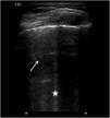
A 6-year-old boy subjected to Fontan surgery, with failure following extubation due to plastic bronchitis (PB). After four days the patient presented hypophonesis of the right upper lobe (RUL). Lung ultrasound evidenced A-lines (asterisk) and some B-lines (white dot), with lung pulse but no lung sliding and no condensation zones. The rest of the lung was well aerated, with lung sliding and no pathological images. Pneumothorax was discarded (Fig. 1), though the absence of lung sliding drew our attention. An increase in density of the RUL was noted after three days (Fig. 2), with no associated clinical findings. After 15 days the patient developed progressive acute respiratory failure, with improved aeration of the right side of the chest at radiography (Fig. 3A), though collapse was recorded after 7 h (Fig. 3B). A new PB cast was extracted via rigid bronchoscopy (Fig. 3C). Lung ultrasound allows the early identification of poorly aerated areas without lung sliding, prior to consolidation. This image could constitute an early indicator of PB in patients at risk of presenting the disorder, and serial lung ultrasound studies could facilitate its evolutive control.
Financial supportThe authors declare that there are no relevant financial or non-financial aspects related to the present study.
Please cite this article as: Bobillo-Perez S, Balaguer M, Cambra FJ. Ecografía pulmonar en bronquitis plástica. Med Intensiva. 2022;46:116–117.


