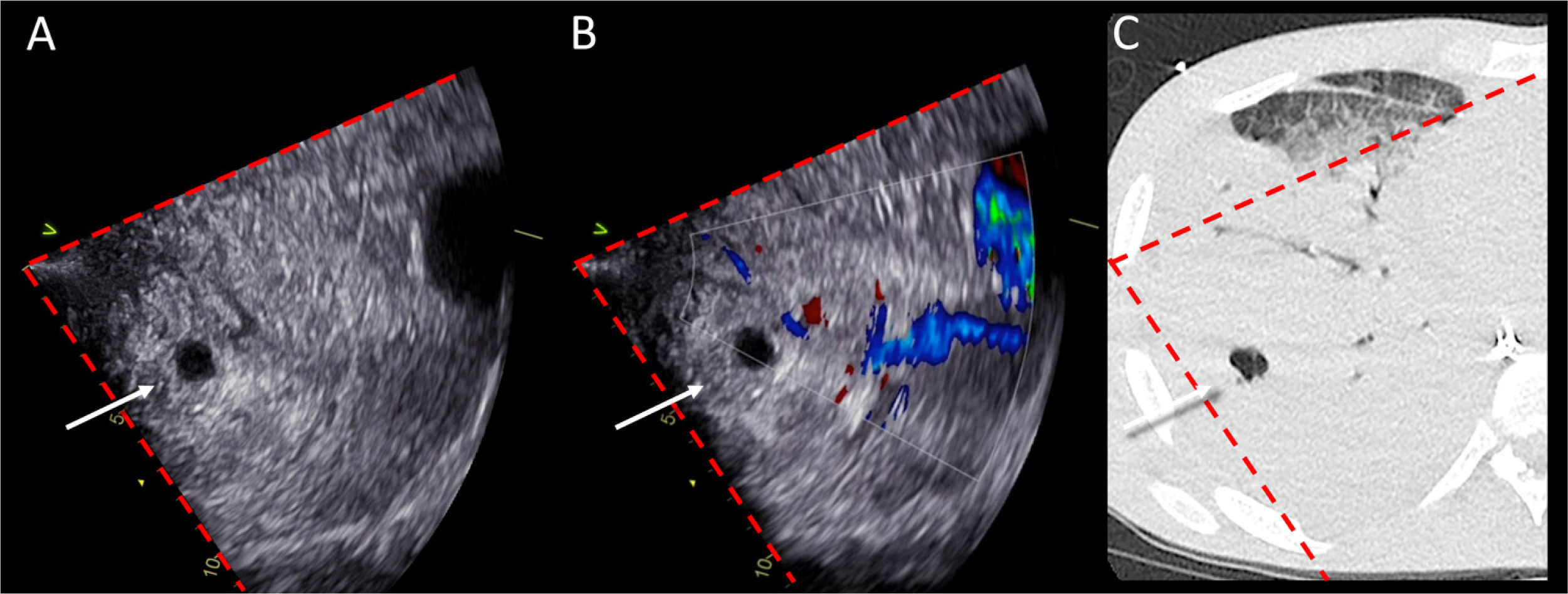

Lung ultrasound was used to monitor a patient admitted to ICU for acute respiratory distress syndrome secondary to Influenza A, requiring mechanical ventilation and veno-venous extra-corporeal membrane oxygenation for refractory hypoxemia. ICU stay was complicated by a Staphylococcus aureus ventilator-associated pneumonia. Figure 1: Panel A shows a small anechoic round image (white arrow) within a tissue-like pattern, compatible with both a pulmonary vessel in short axis and a lung abscess within a consolidated parenchyma (Video 1). A second larger anechoic round image is visualized in deeper fields. Color Doppler (Panel B) easily allows distinguishing abscess as non-pulsatile structures (Video 2). On the corresponding CT scan (Panel C), the red dotted lines delineate the ultrasound beam. Ultrasound well identifies lung abscesses within consolidations as round well-defined anechoic images with posterior enhancement and no pulsatility at colour Doppler; this simple bedside application avoids the need of traditional radiology and allows dynamic guidance to procedures, as needle aspiration.
Impact factor
The Impact Factor measures the average number of citations received in a particular year by papers published in the journal during the two preceding years.
© Clarivate Analytics, Journal Citation Reports 2025
Impact factor 2024
3.1
Citescore 2024
2.3
SJR
SRJ is a prestige metric based on the idea that not all citations are the same. SJR uses a similar algorithm as the Google page rank; it provides a quantitative and qualitative measure of the journal's impact.
See moreSJR 2024
0.353
SNIP
SNIP measures contextual citation impact by wighting citations based on the total number of citations in a subject field.
See moreSNIP 2024
0.573
View more metrics