


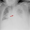
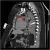
Although echography guidance can increase the success rate of central venous catheter (CVC) placement, it only ensures that the catheter is correctly placed at the level of the vessel entrance. Once the guidewire is inserted there is little we can do to ensure its correct direction on a simple procedure like this one. This is the example of this patient, with left pneumonic empyema in which x-ray confirmation showed CVC migration (Fig. 1). Anecdotally, the only adverse effect reported from this misplacement was acute chest pain on fluids bolus infusion. On chest computed tomography scan, the catheter can be seen going down the left jugular vein and straight inside the brachiocephalic vein (Fig. 2 star) toward the left internal thoracic vein (Fig. 2 arrow).


This incident highlights the importance of routine X-ray visualization of CVC placement, as echography alone cannot completely replace it.

