array:23 [
  "pii" => "S0210569120303375"
  "issn" => "02105691"
  "doi" => "10.1016/j.medin.2020.10.004"
  "estado" => "S300"
  "fechaPublicacion" => "2023-02-01"
  "aid" => "1602"
  "copyright" => "Elsevier España, S.L.U. y SEMICYUC"
  "copyrightAnyo" => "2020"
  "documento" => "article"
  "crossmark" => 1
  "subdocumento" => "sco"
  "cita" => "Med Intensiva. 2023;47:122-3"
  "abierto" => array:3 [
    "ES" => true
    "ES2" => true
    "LATM" => true
  ]
  "gratuito" => true
  "lecturas" => array:1 [
    "total" => 0
  ]
  "itemSiguiente" => array:19 [
    "pii" => "S0210569121000024"
    "issn" => "02105691"
    "doi" => "10.1016/j.medin.2020.12.003"
    "estado" => "S300"
    "fechaPublicacion" => "2023-02-01"
    "aid" => "1615"
    "copyright" => "Elsevier España, S.L.U. y SEMICYUC"
    "documento" => "article"
    "crossmark" => 1
    "subdocumento" => "sco"
    "cita" => "Med Intensiva. 2023;47:124"
    "abierto" => array:3 [
      "ES" => true
      "ES2" => true
      "LATM" => true
    ]
    "gratuito" => true
    "lecturas" => array:1 [
      "total" => 0
    ]
    "es" => array:11 [
      "idiomaDefecto" => true
      "cabecera" => "<span class="elsevierStyleTextfn">Im&#225;genes en Medicina Intensiva</span>"
      "titulo" => "Diagn&#243;stico de neumon&#237;a cavitada por ecograf&#237;a pulmonar"
      "tienePdf" => "es"
      "tieneTextoCompleto" => "es"
      "paginas" => array:1 [
        0 => array:1 [
          "paginaInicial" => "124"
        ]
      ]
      "titulosAlternativos" => array:1 [
        "en" => array:1 [
          "titulo" => "Diagnosis of cavitated pneumonia by lung ultrasound"
        ]
      ]
      "contieneTextoCompleto" => array:1 [
        "es" => true
      ]
      "contienePdf" => array:1 [
        "es" => true
      ]
      "resumenGrafico" => array:2 [
        "original" => 0
        "multimedia" => array:6 [
          "identificador" => "fig0005"
          "etiqueta" => "Figura 1"
          "tipo" => "MULTIMEDIAFIGURA"
          "mostrarFloat" => true
          "mostrarDisplay" => false
          "figura" => array:1 [
            0 => array:4 [
              "imagen" => "gr1.jpeg"
              "Alto" => 488
              "Ancho" => 1874
              "Tamanyo" => 134603
            ]
          ]
        ]
      ]
      "autores" => array:1 [
        0 => array:2 [
          "autoresLista" => "A&#46; G&#243;mez Carranza, A&#46; Santos Peral, R&#46; Vicho Pereira"
          "autores" => array:3 [
            0 => array:2 [
              "nombre" => "A&#46;"
              "apellidos" => "G&#243;mez Carranza"
            ]
            1 => array:2 [
              "nombre" => "A&#46;"
              "apellidos" => "Santos Peral"
            ]
            2 => array:2 [
              "nombre" => "R&#46;"
              "apellidos" => "Vicho Pereira"
            ]
          ]
        ]
      ]
    ]
    "idiomaDefecto" => "es"
    "Traduccion" => array:1 [
      "en" => array:9 [
        "pii" => "S2173572722002946"
        "doi" => "10.1016/j.medine.2022.10.010"
        "estado" => "S300"
        "subdocumento" => ""
        "abierto" => array:3 [
          "ES" => true
          "ES2" => true
          "LATM" => true
        ]
        "gratuito" => true
        "lecturas" => array:1 [
          "total" => 0
        ]
        "idiomaDefecto" => "en"
        "EPUB" => "https://multimedia.elsevier.es/PublicationsMultimediaV1/item/epub/S2173572722002946?idApp=WMIE"
      ]
    ]
    "EPUB" => "https://multimedia.elsevier.es/PublicationsMultimediaV1/item/epub/S0210569121000024?idApp=WMIE"
    "url" => "/02105691/0000004700000002/v1_202301260905/S0210569121000024/v1_202301260905/es/main.assets"
  ]
  "itemAnterior" => array:18 [
    "pii" => "S0210569120303211"
    "issn" => "02105691"
    "doi" => "10.1016/j.medin.2020.09.005"
    "estado" => "S300"
    "fechaPublicacion" => "2023-02-01"
    "aid" => "1592"
    "copyright" => "Elsevier Espa&#241;a&#44; S&#46;L&#46;U&#46; y SEMICYUC"
    "documento" => "article"
    "crossmark" => 1
    "subdocumento" => "sco"
    "cita" => "Med Intensiva. 2023;47:120-1"
    "abierto" => array:3 [
      "ES" => true
      "ES2" => true
      "LATM" => true
    ]
    "gratuito" => true
    "lecturas" => array:1 [
      "total" => 0
    ]
    "es" => array:11 [
      "idiomaDefecto" => true
      "cabecera" => "<span class="elsevierStyleTextfn">Im&#225;genes en Medicina Intensiva</span>"
      "titulo" => "Diagn&#243;stico de par&#225;lisis diafragm&#225;tica tras cirug&#237;a cardiaca mediante ecograf&#237;a pulmonar"
      "tienePdf" => "es"
      "tieneTextoCompleto" => "es"
      "paginas" => array:1 [
        0 => array:2 [
          "paginaInicial" => "120"
          "paginaFinal" => "121"
        ]
      ]
      "titulosAlternativos" => array:1 [
        "en" => array:1 [
          "titulo" => "Diagnosis of diaphragmatic paralysis by ultrasound after cardiac surgery"
        ]
      ]
      "contieneTextoCompleto" => array:1 [
        "es" => true
      ]
      "contienePdf" => array:1 [
        "es" => true
      ]
      "resumenGrafico" => array:2 [
        "original" => 0
        "multimedia" => array:6 [
          "identificador" => "fig0005"
          "etiqueta" => "Figura 1"
          "tipo" => "MULTIMEDIAFIGURA"
          "mostrarFloat" => true
          "mostrarDisplay" => false
          "figura" => array:1 [
            0 => array:4 [
              "imagen" => "gr1.jpeg"
              "Alto" => 1364
              "Ancho" => 1267
              "Tamanyo" => 311455
            ]
          ]
        ]
      ]
      "autores" => array:1 [
        0 => array:2 [
          "autoresLista" => "A&#46; Santos Peral, A&#46; G&#243;mez Carranza, R&#46; Vicho Pereira"
          "autores" => array:3 [
            0 => array:2 [
              "nombre" => "A&#46;"
              "apellidos" => "Santos Peral"
            ]
            1 => array:2 [
              "nombre" => "A&#46;"
              "apellidos" => "G&#243;mez Carranza"
            ]
            2 => array:2 [
              "nombre" => "R&#46;"
              "apellidos" => "Vicho Pereira"
            ]
          ]
        ]
      ]
    ]
    "idiomaDefecto" => "es"
    "EPUB" => "https://multimedia.elsevier.es/PublicationsMultimediaV1/item/epub/S0210569120303211?idApp=WMIE"
    "url" => "/02105691/0000004700000002/v1_202301260905/S0210569120303211/v1_202301260905/es/main.assets"
  ]
  "en" => array:13 [
    "idiomaDefecto" => true
    "cabecera" => "<span class="elsevierStyleTextfn">Images in Intensive Medicine</span>"
    "titulo" => "Jump to the diagnosis from Hampton&#39;s hump"
    "tieneTextoCompleto" => true
    "paginas" => array:1 [
      0 => array:2 [
        "paginaInicial" => "122"
        "paginaFinal" => "123"
      ]
    ]
    "autores" => array:1 [
      0 => array:4 [
        "autoresLista" => "L&#46;-T&#46; Keng, H&#46;-Y&#46; Pan"
        "autores" => array:2 [
          0 => array:4 [
            "nombre" => "L&#46;-T&#46;"
            "apellidos" => "Keng"
            "email" => array:1 [
              0 => "ltkeng@gmail.com"
            ]
            "referencia" => array:1 [
              0 => array:2 [
                "etiqueta" => "<span class="elsevierStyleSup">&#42;</span>"
                "identificador" => "cor0005"
              ]
            ]
          ]
          1 => array:2 [
            "nombre" => "H&#46;-Y&#46;"
            "apellidos" => "Pan"
          ]
        ]
        "afiliaciones" => array:1 [
          0 => array:2 [
            "entidad" => "Department of Internal Medicine&#44; National Taiwan University Hospital Hsin-Chu Branch&#44; Hsin-Chu&#44; Taiwan"
            "identificador" => "aff0005"
          ]
        ]
        "correspondencia" => array:1 [
          0 => array:3 [
            "identificador" => "cor0005"
            "etiqueta" => "&#8270;"
            "correspondencia" => "Corresponding author&#46;"
          ]
        ]
      ]
    ]
    "titulosAlternativos" => array:1 [
      "es" => array:1 [
        "titulo" => "Ir al diagn&#243;stico de la joroba de hampton"
      ]
    ]
    "resumenGrafico" => array:2 [
      "original" => 0
      "multimedia" => array:6 [
        "identificador" => "fig0010"
        "etiqueta" => "Figure 1"
        "tipo" => "MULTIMEDIAFIGURA"
        "mostrarFloat" => true
        "mostrarDisplay" => false
        "figura" => array:1 [
          0 => array:4 [
            "imagen" => "gr1.jpeg"
            "Alto" => 1330
            "Ancho" => 1200
            "Tamanyo" => 136852
          ]
        ]
      ]
    ]
    "textoCompleto" => "<span class="elsevierStyleSections"><p id="par0010" class="elsevierStylePara elsevierViewall">A 78-year-old bedridden man was admitted for presumed aspiration pneumonia with a chest radiograph showing a patchy opacity in the right upper lung zone &#40;<a class="elsevierStyleCrossRef" href="#fig0010">Fig&#46; 1</a>&#41;&#44; with the presentation of fever and dyspnea&#46; An urgent CAT scan revealed a wedge-shaped opacity in the right upper lobe &#40;<a class="elsevierStyleCrossRef" href="#fig0015">Fig&#46; 2</a>&#41; and filling defects in the distal right main pulmonary artery &#40;<a class="elsevierStyleCrossRef" href="#fig0020">Fig&#46; 3</a>&#41;&#44; confirming the diagnosis of pulmonary embolism with distal pulmonary infarction &#40;Hampton&#39;s hump&#41;&#46; Further examination revealed a deep vein thrombosis involving both legs&#46; He was successfully treated with anticoagulant therapy&#46;</p><elsevierMultimedia ident="fig0010"></elsevierMultimedia><elsevierMultimedia ident="fig0015"></elsevierMultimedia><elsevierMultimedia ident="fig0020"></elsevierMultimedia><p id="par0015" class="elsevierStylePara elsevierViewall">The presence of high fever and radiographic pulmonary opacity may be compatible with pneumonia&#46; In patients with risk factors for venous thromboembolism&#44; only vigilant detection of the Hampton&#39;s hump on chest radiography could jump to the correct diagnosis of acute pulmonary embolism&#46;</p><span id="sec0005" class="elsevierStyleSection elsevierViewall"><span class="elsevierStyleSectionTitle" id="sect0005">Authors&#8217; contributors</span><p id="par0020" class="elsevierStylePara elsevierViewall">Li-Ta Keng drafted the manuscript&#46; Heng-Yu Pan prepared the image and revised the manuscript&#46;</p></span><span id="sec0015" class="elsevierStyleSection elsevierViewall"><span class="elsevierStyleSectionTitle" id="sect0010">Patient consent</span><p id="par0030" class="elsevierStylePara elsevierViewall">Obtained&#46;</p></span><span id="sec0010" class="elsevierStyleSection elsevierViewall"><span class="elsevierStyleSectionTitle" id="sect0015">Financial support</span><p id="par0035" class="elsevierStylePara elsevierViewall">None declared&#46;</p></span><span id="sec0020" class="elsevierStyleSection elsevierViewall"><span class="elsevierStyleSectionTitle" id="sect0020">Conflict of interest</span><p id="par0025" class="elsevierStylePara elsevierViewall">None declared&#46;</p></span></span>"
    "textoCompletoSecciones" => array:1 [
      "secciones" => array:4 [
        0 => array:2 [
          "identificador" => "sec0005"
          "titulo" => "Authors&#8217; contributors"
        ]
        1 => array:2 [
          "identificador" => "sec0015"
          "titulo" => "Patient consent"
        ]
        2 => array:2 [
          "identificador" => "sec0010"
          "titulo" => "Financial support"
        ]
        3 => array:2 [
          "identificador" => "sec0020"
          "titulo" => "Conflict of interest"
        ]
      ]
    ]
    "pdfFichero" => "main.pdf"
    "tienePdf" => true
    "multimedia" => array:3 [
      0 => array:6 [
        "identificador" => "fig0010"
        "etiqueta" => "Figure 1"
        "tipo" => "MULTIMEDIAFIGURA"
        "mostrarFloat" => true
        "mostrarDisplay" => false
        "figura" => array:1 [
          0 => array:4 [
            "imagen" => "gr1.jpeg"
            "Alto" => 1330
            "Ancho" => 1200
            "Tamanyo" => 136852
          ]
        ]
      ]
      1 => array:6 [
        "identificador" => "fig0015"
        "etiqueta" => "Figure 2"
        "tipo" => "MULTIMEDIAFIGURA"
        "mostrarFloat" => true
        "mostrarDisplay" => false
        "figura" => array:1 [
          0 => array:4 [
            "imagen" => "gr2.jpeg"
            "Alto" => 868
            "Ancho" => 900
            "Tamanyo" => 65258
          ]
        ]
      ]
      2 => array:6 [
        "identificador" => "fig0020"
        "etiqueta" => "Figure 3"
        "tipo" => "MULTIMEDIAFIGURA"
        "mostrarFloat" => true
        "mostrarDisplay" => false
        "figura" => array:1 [
          0 => array:4 [
            "imagen" => "gr3.jpeg"
            "Alto" => 869
            "Ancho" => 900
            "Tamanyo" => 80854
          ]
        ]
      ]
    ]
  ]
  "idiomaDefecto" => "en"
  "url" => "/02105691/0000004700000002/v1_202301260905/S0210569120303375/v1_202301260905/en/main.assets"
  "Apartado" => array:4 [
    "identificador" => "440"
    "tipo" => "SECCION"
    "es" => array:2 [
      "titulo" => "Im&#225;genes en medicina intensiva"
      "idiomaDefecto" => true
    ]
    "idiomaDefecto" => "es"
  ]
  "PDF" => "https://static.elsevier.es/multimedia/02105691/0000004700000002/v1_202301260905/S0210569120303375/v1_202301260905/en/main.pdf?idApp=WMIE&text.app=https://medintensiva.org/"
  "EPUB" => "https://multimedia.elsevier.es/PublicationsMultimediaV1/item/epub/S0210569120303375?idApp=WMIE"
]
Compartir
Información de la revista
Vol. 47. Núm. 2.
Páginas 122-123 (febrero 2023)
Compartir
Compartir
Descargar PDF
Más opciones de artículo
Visitas
1937
Vol. 47. Núm. 2.
Páginas 122-123 (febrero 2023)
Images in Intensive Medicine
Acceso a texto completo
Jump to the diagnosis from Hampton's hump
Ir al diagnóstico de la joroba de hampton
Visitas
1937
L.-T. Keng
Autor para correspondencia
ltkeng@gmail.com

Corresponding author.
, H.-Y. Pan
Department of Internal Medicine, National Taiwan University Hospital Hsin-Chu Branch, Hsin-Chu, Taiwan
Este artículo ha recibido
Información del artículo
Texto completo
Descargar PDF
Estadísticas
Figuras (3)
Mostrar másMostrar menos
Texto completo

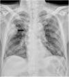
A 78-year-old bedridden man was admitted for presumed aspiration pneumonia with a chest radiograph showing a patchy opacity in the right upper lung zone (Fig. 1), with the presentation of fever and dyspnea. An urgent CAT scan revealed a wedge-shaped opacity in the right upper lobe (Fig. 2) and filling defects in the distal right main pulmonary artery (Fig. 3), confirming the diagnosis of pulmonary embolism with distal pulmonary infarction (Hampton's hump). Further examination revealed a deep vein thrombosis involving both legs. He was successfully treated with anticoagulant therapy.

Figure 1
(0.13MB).
Figure 2
(0.06MB).
Figure 3
(0.08MB).

The presence of high fever and radiographic pulmonary opacity may be compatible with pneumonia. In patients with risk factors for venous thromboembolism, only vigilant detection of the Hampton's hump on chest radiography could jump to the correct diagnosis of acute pulmonary embolism.

Authors’ contributors

Li-Ta Keng drafted the manuscript. Heng-Yu Pan prepared the image and revised the manuscript.

Patient consent

Obtained.

Financial support

None declared.

Conflict of interest

None declared.

Copyright © 2020. Elsevier España, S.L.U. y SEMICYUC
Descargar PDF
Idiomas
Medicina Intensiva
Opciones de artículo
Herramientas
es en

¿Es usted profesional sanitario apto para prescribir o dispensar medicamentos?

Are you a health professional able to prescribe or dispense drugs?