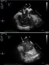
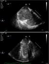
A 44-year-old man was admitted to our ICU due to thoracic trauma with associated bilateral pulmonary contusion. He developed severe refractory respiratory failure (PaO2/FiO2<100) and hemodynamic instability secondary to hypoxemia. Due to the inability to oxygenate the patient, it was decided to begin with veno-venous ECMO therapy. During cannulation of the femoral vein, a large thrombus (white arrow) was observed in the right atrium passing through the tricuspid valve into the pulmonary artery (Fig. 1); suddenly, the patient suffered cardiac arrest in pulseless electrical activity (Fig. 2), requiring advanced CPR maneuvers, recovering spontaneous circulation once ECMO therapy begun (Video). He remained under ECMO for seven days, after which, it was withdrawn due to respiratory improvement. He presented full neurological recovery.
array:23 [ "pii" => "S0210569118303073" "issn" => "02105691" "doi" => "10.1016/j.medin.2018.10.003" "estado" => "S300" "fechaPublicacion" => "2019-08-01" "aid" => "1287" "copyright" => "Elsevier España, S.L.U. y SEMICYUC" "copyrightAnyo" => "2018" "documento" => "article" "crossmark" => 1 "subdocumento" => "sco" "cita" => "Med Intensiva. 2019;43:390-1" "abierto" => array:3 [ "ES" => true "ES2" => true "LATM" => true ] "gratuito" => true "lecturas" => array:2 [ "total" => 340 "formatos" => array:3 [ "EPUB" => 57 "HTML" => 187 "PDF" => 96 ] ] "itemSiguiente" => array:19 [ "pii" => "S0210569118301918" "issn" => "02105691" "doi" => "10.1016/j.medin.2018.06.007" "estado" => "S300" "fechaPublicacion" => "2019-08-01" "aid" => "1236" "copyright" => "Elsevier España, S.L.U. y SEMICYUC" "documento" => "article" "crossmark" => 1 "subdocumento" => "sco" "cita" => "Med Intensiva. 2019;43:392" "abierto" => array:3 [ "ES" => true "ES2" => true "LATM" => true ] "gratuito" => true "lecturas" => array:2 [ "total" => 780 "formatos" => array:3 [ "EPUB" => 72 "HTML" => 550 "PDF" => 158 ] ] "es" => array:11 [ "idiomaDefecto" => true "cabecera" => "<span class="elsevierStyleTextfn">Imágenes en Medicina Intensiva</span>" "titulo" => "SCIWORA, una lesión a conocer y a perseguir" "tienePdf" => "es" "tieneTextoCompleto" => "es" "paginas" => array:1 [ 0 => array:1 [ "paginaInicial" => "392" ] ] "titulosAlternativos" => array:1 [ "en" => array:1 [ "titulo" => "SCIWORA, an injury to know and diagnose" ] ] "contieneTextoCompleto" => array:1 [ "es" => true ] "contienePdf" => array:1 [ "es" => true ] "resumenGrafico" => array:2 [ "original" => 0 "multimedia" => array:7 [ "identificador" => "fig0005" "etiqueta" => "Figura 1" "tipo" => "MULTIMEDIAFIGURA" "mostrarFloat" => true "mostrarDisplay" => false "figura" => array:1 [ 0 => array:4 [ "imagen" => "gr1.jpeg" "Alto" => 1000 "Ancho" => 1000 "Tamanyo" => 134262 ] ] "descripcion" => array:1 [ "es" => "<p id="spar0005" class="elsevierStyleSimplePara elsevierViewall">A) Tomografía computarizada (TC) de 2008: corte axial a nivel de C1-C2 con estenosis de canal medular de 6<span class="elsevierStyleHsp" style=""></span>mm (asterisco gris). B) Corte axial a nivel de C1 con estenosis de canal medular en TC en 2018 de 6<span class="elsevierStyleHsp" style=""></span>mm (asterisco gris). C) Corte sagital en TC en la que se objetiva estenosis de canal en unión craneocervical ya conocida sin lesión aparente de partes blandas (flecha gris sin relleno). D) Resonancia magnética en secuencia T1: estenosis raquídea con engrosamiento dural difuso y alteración de la señal en región inferior de bulbo, así como intramedular cervical compatible con mielopatía traumática (flecha gris con relleno).</p>" ] ] ] "autores" => array:1 [ 0 => array:2 [ "autoresLista" => "R. Viejo Moreno, C. Benito Puncel, Z. Eguileor Marín" "autores" => array:3 [ 0 => array:2 [ "nombre" => "R." "apellidos" => "Viejo Moreno" ] 1 => array:2 [ "nombre" => "C." "apellidos" => "Benito Puncel" ] 2 => array:2 [ "nombre" => "Z." "apellidos" => "Eguileor Marín" ] ] ] ] ] "idiomaDefecto" => "es" "Traduccion" => array:1 [ "en" => array:9 [ "pii" => "S2173572719300980" "doi" => "10.1016/j.medine.2019.05.006" "estado" => "S300" "subdocumento" => "" "abierto" => array:3 [ "ES" => true "ES2" => true "LATM" => true ] "gratuito" => true "lecturas" => array:1 [ "total" => 0 ] "idiomaDefecto" => "en" "EPUB" => "https://multimedia.elsevier.es/PublicationsMultimediaV1/item/epub/S2173572719300980?idApp=WMIE" ] ] "EPUB" => "https://multimedia.elsevier.es/PublicationsMultimediaV1/item/epub/S0210569118301918?idApp=WMIE" "url" => "/02105691/0000004300000006/v1_201907230742/S0210569118301918/v1_201907230742/es/main.assets" ] "itemAnterior" => array:18 [ "pii" => "S0210569118303097" "issn" => "02105691" "doi" => "10.1016/j.medin.2018.10.005" "estado" => "S300" "fechaPublicacion" => "2019-08-01" "aid" => "1289" "documento" => "article" "crossmark" => 1 "subdocumento" => "sco" "cita" => "Med Intensiva. 2019;43:389" "abierto" => array:3 [ "ES" => true "ES2" => true "LATM" => true ] "gratuito" => true "lecturas" => array:2 [ "total" => 381 "formatos" => array:3 [ "EPUB" => 83 "HTML" => 188 "PDF" => 110 ] ] "es" => array:11 [ "idiomaDefecto" => true "cabecera" => "<span class="elsevierStyleTextfn">Imágenes en Medicina Intensiva</span>" "titulo" => "Sangrado de arteria mamaria interna secundario a resucitación prolongada" "tienePdf" => "es" "tieneTextoCompleto" => "es" "paginas" => array:1 [ 0 => array:1 [ "paginaInicial" => "389" ] ] "titulosAlternativos" => array:1 [ "en" => array:1 [ "titulo" => "Bleeding from internal mammary artery related to prolonged cardiopulmonary resuscitation" ] ] "contieneTextoCompleto" => array:1 [ "es" => true ] "contienePdf" => array:1 [ "es" => true ] "resumenGrafico" => array:2 [ "original" => 0 "multimedia" => array:6 [ "identificador" => "fig0005" "etiqueta" => "Figura 1" "tipo" => "MULTIMEDIAFIGURA" "mostrarFloat" => true "mostrarDisplay" => false "figura" => array:1 [ 0 => array:4 [ "imagen" => "gr1.jpeg" "Alto" => 722 "Ancho" => 819 "Tamanyo" => 84267 ] ] ] ] "autores" => array:1 [ 0 => array:2 [ "autoresLista" => "V. Arnaiz Arnaiz, C. Berástegi Santamaría, P.M. Olaechea Astigarraga" "autores" => array:3 [ 0 => array:2 [ "nombre" => "V." "apellidos" => "Arnaiz Arnaiz" ] 1 => array:2 [ "nombre" => "C." "apellidos" => "Berástegi Santamaría" ] 2 => array:2 [ "nombre" => "P.M." "apellidos" => "Olaechea Astigarraga" ] ] ] ] ] "idiomaDefecto" => "es" "Traduccion" => array:1 [ "en" => array:9 [ "pii" => "S2173572719300967" "doi" => "10.1016/j.medine.2019.05.004" "estado" => "S300" "subdocumento" => "" "abierto" => array:3 [ "ES" => true "ES2" => true "LATM" => true ] "gratuito" => true "lecturas" => array:1 [ "total" => 0 ] "idiomaDefecto" => "en" "EPUB" => "https://multimedia.elsevier.es/PublicationsMultimediaV1/item/epub/S2173572719300967?idApp=WMIE" ] ] "EPUB" => "https://multimedia.elsevier.es/PublicationsMultimediaV1/item/epub/S0210569118303097?idApp=WMIE" "url" => "/02105691/0000004300000006/v1_201907230742/S0210569118303097/v1_201907230742/es/main.assets" ] "en" => array:13 [ "idiomaDefecto" => true "cabecera" => "<span class="elsevierStyleTextfn">Image in Intensive Medicine</span>" "titulo" => "Mobile thrombus in right atrium during cannulation of veno-venous extracorporeal membrane oxygenation" "tieneTextoCompleto" => true "paginas" => array:1 [ 0 => array:2 [ "paginaInicial" => "390" "paginaFinal" => "391" ] ] "autores" => array:1 [ 0 => array:4 [ "autoresLista" => "E. Rodriguez-Ruiz, A. López-Lago, M. Álvarez Barredo" "autores" => array:3 [ 0 => array:4 [ "nombre" => "E." "apellidos" => "Rodriguez-Ruiz" "email" => array:1 [ 0 => "r.ruizemilio@gmail.com" ] "referencia" => array:3 [ 0 => array:2 [ "etiqueta" => "<span class="elsevierStyleSup">a</span>" "identificador" => "aff0005" ] 1 => array:2 [ "etiqueta" => "<span class="elsevierStyleSup">b</span>" "identificador" => "aff0010" ] 2 => array:2 [ "etiqueta" => "<span class="elsevierStyleSup">*</span>" "identificador" => "cor0005" ] ] ] 1 => array:3 [ "nombre" => "A." "apellidos" => "López-Lago" "referencia" => array:2 [ 0 => array:2 [ "etiqueta" => "<span class="elsevierStyleSup">a</span>" "identificador" => "aff0005" ] 1 => array:2 [ "etiqueta" => "<span class="elsevierStyleSup">b</span>" "identificador" => "aff0010" ] ] ] 2 => array:3 [ "nombre" => "M." "apellidos" => "Álvarez Barredo" "referencia" => array:1 [ 0 => array:2 [ "etiqueta" => "<span class="elsevierStyleSup">c</span>" "identificador" => "aff0015" ] ] ] ] "afiliaciones" => array:3 [ 0 => array:3 [ "entidad" => "Intensive Care Medicine Department, University Clinical Hospital of Santiago de Compostela, SERGAS, Spain" "etiqueta" => "a" "identificador" => "aff0005" ] 1 => array:3 [ "entidad" => "University of Santiago de Compostela, Santiago de Compostela, A Coruña, Spain" "etiqueta" => "b" "identificador" => "aff0010" ] 2 => array:3 [ "entidad" => "Cardiology Department, University Clinical Hospital of Santiago de Compostela, Spain" "etiqueta" => "c" "identificador" => "aff0015" ] ] "correspondencia" => array:1 [ 0 => array:3 [ "identificador" => "cor0005" "etiqueta" => "⁎" "correspondencia" => "Corresponding author." ] ] ] ] "titulosAlternativos" => array:1 [ "es" => array:1 [ "titulo" => "Trombo móvil en la aurícula derecha durante la canulación de una membrana de oxigenación estracorpórea veno-venosa" ] ] "resumenGrafico" => array:2 [ "original" => 0 "multimedia" => array:6 [ "identificador" => "fig0005" "etiqueta" => "Figure 1" "tipo" => "MULTIMEDIAFIGURA" "mostrarFloat" => true "mostrarDisplay" => false "figura" => array:1 [ 0 => array:4 [ "imagen" => "gr1.jpeg" "Alto" => 1980 "Ancho" => 1500 "Tamanyo" => 206826 ] ] ] ] "textoCompleto" => "<span class="elsevierStyleSections"><p id="par0005" class="elsevierStylePara elsevierViewall">A 44-year-old man was admitted to our ICU due to thoracic trauma with associated bilateral pulmonary contusion. He developed severe refractory respiratory failure (PaO2/FiO2<span class="elsevierStyleHsp" style=""></span><<span class="elsevierStyleHsp" style=""></span>100) and hemodynamic instability secondary to hypoxemia. Due to the inability to oxygenate the patient, it was decided to begin with veno-venous ECMO therapy. During cannulation of the femoral vein, a large thrombus (white arrow) was observed in the right atrium passing through the tricuspid valve into the pulmonary artery (<a class="elsevierStyleCrossRef" href="#fig0005">Fig. 1</a>); suddenly, the patient suffered cardiac arrest in pulseless electrical activity (<a class="elsevierStyleCrossRef" href="#fig0010">Fig. 2</a>), requiring advanced CPR maneuvers, recovering spontaneous circulation once ECMO therapy begun (Video). He remained under ECMO for seven days, after which, it was withdrawn due to respiratory improvement. He presented full neurological recovery.</p><elsevierMultimedia ident="fig0005"></elsevierMultimedia><elsevierMultimedia ident="fig0010"></elsevierMultimedia></span>" "pdfFichero" => "main.pdf" "tienePdf" => true "apendice" => array:1 [ 0 => array:1 [ "seccion" => array:1 [ 0 => array:4 [ "apendice" => "<p id="par0015" class="elsevierStylePara elsevierViewall">The following are the supplementary data to this article:<elsevierMultimedia ident="upi0005"></elsevierMultimedia></p>" "etiqueta" => "Appendix A" "titulo" => "Supplementary data" "identificador" => "sec0010" ] ] ] ] "multimedia" => array:3 [ 0 => array:6 [ "identificador" => "fig0005" "etiqueta" => "Figure 1" "tipo" => "MULTIMEDIAFIGURA" "mostrarFloat" => true "mostrarDisplay" => false "figura" => array:1 [ 0 => array:4 [ "imagen" => "gr1.jpeg" "Alto" => 1980 "Ancho" => 1500 "Tamanyo" => 206826 ] ] ] 1 => array:6 [ "identificador" => "fig0010" "etiqueta" => "Figure 2" "tipo" => "MULTIMEDIAFIGURA" "mostrarFloat" => true "mostrarDisplay" => false "figura" => array:1 [ 0 => array:4 [ "imagen" => "gr2.jpeg" "Alto" => 1971 "Ancho" => 1500 "Tamanyo" => 210284 ] ] ] 2 => array:5 [ "identificador" => "upi0005" "tipo" => "MULTIMEDIAECOMPONENTE" "mostrarFloat" => false "mostrarDisplay" => true "Ecomponente" => array:3 [ "fichero" => "mmc1.mp4" "ficheroTamanyo" => 27876860 "Video" => array:2 [ "flv" => array:5 [ "fichero" => "mmc1.flv" "poster" => "mmc1.jpg" "tiempo" => 0 "alto" => 0 "ancho" => 0 ] "mp4" => array:5 [ "fichero" => "mmc1.m4v" "poster" => "mmc1.jpg" "tiempo" => 0 "alto" => 0 "ancho" => 0 ] ] ] ] ] ] "idiomaDefecto" => "en" "url" => "/02105691/0000004300000006/v1_201907230742/S0210569118303073/v1_201907230742/en/main.assets" "Apartado" => array:4 [ "identificador" => "440" "tipo" => "SECCION" "es" => array:2 [ "titulo" => "Imágenes en medicina intensiva" "idiomaDefecto" => true ] "idiomaDefecto" => "es" ] "PDF" => "https://static.elsevier.es/multimedia/02105691/0000004300000006/v1_201907230742/S0210569118303073/v1_201907230742/en/main.pdf?idApp=WMIE&text.app=https://medintensiva.org/" "EPUB" => "https://multimedia.elsevier.es/PublicationsMultimediaV1/item/epub/S0210569118303073?idApp=WMIE" ]