


New-born infants with left congenital diaphragmatic hernia require therapy with veno-venous extracorporeal membrane oxygenation (ECMO) due to pulmonary hypertensive crisis. Forty-eight hours after receiving ECMO therapy, the patient's respiratory parameters get worse and a diagnosis of left lung atelectasis is confirmed (Fig. 1 A) that resolves successfully through thoracic ultrasound-guided alveolar recruitment maneuvers at the patient's bedside (Fig. 1B and C).
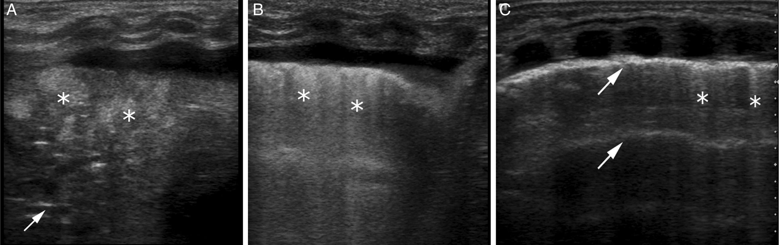
Thoracic ultrasound scans of the patient's left lung. (A) Area of consolidation showing hepatized lungs with aerial bronchogram showing hyperechogenic lines parallel to the pleura in the consolidated area (arrow) and fluid bronchogram with hyperechoic zones in the consolidated area (asterisk), lack of A lines and pleural reinforcement with pleural effusion consistent with a pattern of atelectasis. HFOV respiratory parameters: MAP 12, amplitude 40 and frequency 9. (B) Abundant coalescent B lines (asterisk) indicative of opening of atelectasis in the presence of pulmonary edema. Reinforcement of pleural echogenicity and pleural effusion. Increased respiratory parameters to MAP: 15, amplitude 50 and frequency 8. (C) Lung recruitment with normal parenchyma; presence of A lines (arrow) and scarce B lines (asterisk). Increased respiratory parameters to MAP: 18, amplitude 55 and frequency 8.
Please cite this article as: del Rey Hurtado de Mendoza B, Sanchez-de-Toledo J, Rodríguez-Fanjul J. Ecografía torácica para guiar maniobras de reclutamiento pulmonar. Med Intens. 2018;42:e20.

