


Edited by: Juan Antonio Llompart-Pou - Specialist in Intensive Care Medicine, Doctorate in Health Sciences, Universitat Illes Balears. Department of Intensive Care Medicine, Trauma and Neurocritical ICU, Hospital Universitari Son Espases. Palma, Spain
Last update: November 2025
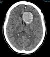
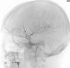
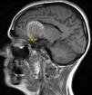
More infoA 49-year-old woman suffered a first seizure episode. The brain CT scan (Fig. 1) revealed a giant aneurysm of the left internal carotid artery (ICA) with subtotal thrombosis (no CT-angio contrast uptake). Arteriography was decided (Fig. 2), showing a large left carotid-ophthalmic aneurysm, with a minimum patent portion (8 × 5 mm). The rest proved thrombotic. During arteriography, the patient developed hemiparesis and deviation of the vocal commissure, with arteriographic vasospasm that subsided with verapamil. Magnetic resonance imaging (Fig. 3) described the aneurysm with a left frontal parasellar location, measuring 4.2 × 3.9 × 3.6 cm, originating at the top of the left ICA and exerting a mass effect upon both anterior cerebral arteries and the left middle cerebral artery, as well as compression and right displacement of both frontal horns. The MR-angio study evidenced a 9-mm saccular dilatation in the supraclinoid portion of the left ICA, located within the lower third of the described lesion. A differential diagnosis with contained rupture of the aneurysm was considered. Surgery with clipping of the aneurysm was decided, with confirmation of its partial thrombosis and an intact capsule. The patient subsequently remained symptoms-free, with salt-wasting syndrome as the only posterior complication.
Financial support


None.
None.
Thanks are due to the Intensive Care Unit of the HUNSC (Santa Cruz de Tenerife) for the training received and to Carmelo for his firm support.

