array:23 [
  "pii" => "S2173572723001807"
  "issn" => "21735727"
  "doi" => "10.1016/j.medine.2023.09.002"
  "estado" => "S300"
  "fechaPublicacion" => "2024-05-01"
  "aid" => "1933"
  "copyright" => "Elsevier España, S.L.U. and SEMICYUC"
  "copyrightAnyo" => "2023"
  "documento" => "article"
  "crossmark" => 1
  "subdocumento" => "sco"
  "cita" => "Med Intensiva. 2024;48:303-4"
  "abierto" => array:3 [
    "ES" => true
    "ES2" => true
    "LATM" => true
  ]
  "gratuito" => true
  "lecturas" => array:1 [
    "total" => 0
  ]
  "itemSiguiente" => array:18 [
    "pii" => "S2173572723002606"
    "issn" => "21735727"
    "doi" => "10.1016/j.medine.2023.12.007"
    "estado" => "S300"
    "fechaPublicacion" => "2024-05-01"
    "aid" => "1963"
    "copyright" => "Elsevier España, S.L.U. and SEMICYUC"
    "documento" => "article"
    "crossmark" => 1
    "subdocumento" => "sco"
    "cita" => "Med Intensiva. 2024;48:305-6"
    "abierto" => array:3 [
      "ES" => true
      "ES2" => true
      "LATM" => true
    ]
    "gratuito" => true
    "lecturas" => array:1 [
      "total" => 0
    ]
    "en" => array:11 [
      "idiomaDefecto" => true
      "cabecera" => "<span class="elsevierStyleTextfn">Images in Intensive Medicine</span>"
      "titulo" => "Point of care lung ultrasound diagnosis of concomitant lung abscess and pleural empyema due to a bronchopleural fistula"
      "tienePdf" => "en"
      "tieneTextoCompleto" => "en"
      "paginas" => array:1 [
        0 => array:2 [
          "paginaInicial" => "305"
          "paginaFinal" => "306"
        ]
      ]
      "titulosAlternativos" => array:1 [
        "es" => array:1 [
          "titulo" => "Diagn&#243;stico ecogr&#225;fico pulmonar de absceso pumonar y empiema pleural concomitantes debido a una f&#237;stula broncopleural"
        ]
      ]
      "contieneTextoCompleto" => array:1 [
        "en" => true
      ]
      "contienePdf" => array:1 [
        "en" => true
      ]
      "resumenGrafico" => array:2 [
        "original" => 0
        "multimedia" => array:7 [
          "identificador" => "fig0005"
          "etiqueta" => "Figure 1"
          "tipo" => "MULTIMEDIAFIGURA"
          "mostrarFloat" => true
          "mostrarDisplay" => false
          "figura" => array:1 [
            0 => array:4 [
              "imagen" => "gr1.jpeg"
              "Alto" => 991
              "Ancho" => 3341
              "Tamanyo" => 292258
            ]
          ]
          "detalles" => array:1 [
            0 => array:3 [
              "identificador" => "at0005"
              "detalle" => "Figure "
              "rol" => "short"
            ]
          ]
        ]
      ]
      "autores" => array:1 [
        0 => array:2 [
          "autoresLista" => "Adrien Rivory, Gary Duclos, Laurent Zieleskiewicz"
          "autores" => array:3 [
            0 => array:2 [
              "nombre" => "Adrien"
              "apellidos" => "Rivory"
            ]
            1 => array:2 [
              "nombre" => "Gary"
              "apellidos" => "Duclos"
            ]
            2 => array:2 [
              "nombre" => "Laurent"
              "apellidos" => "Zieleskiewicz"
            ]
          ]
        ]
      ]
    ]
    "idiomaDefecto" => "en"
    "EPUB" => "https://multimedia.elsevier.es/PublicationsMultimediaV1/item/epub/S2173572723002606?idApp=WMIE"
    "url" => "/21735727/0000004800000005/v1_202404281348/S2173572723002606/v1_202404281348/en/main.assets"
  ]
  "itemAnterior" => array:19 [
    "pii" => "S217357272400016X"
    "issn" => "21735727"
    "doi" => "10.1016/j.medine.2024.01.007"
    "estado" => "S300"
    "fechaPublicacion" => "2024-05-01"
    "aid" => "1973"
    "copyright" => "Elsevier Espa&#241;a&#44; S&#46;L&#46;U&#46; and SEMICYUC"
    "documento" => "simple-article"
    "crossmark" => 1
    "subdocumento" => "cor"
    "cita" => "Med Intensiva. 2024;48:301-2"
    "abierto" => array:3 [
      "ES" => true
      "ES2" => true
      "LATM" => true
    ]
    "gratuito" => true
    "lecturas" => array:1 [
      "total" => 0
    ]
    "en" => array:11 [
      "idiomaDefecto" => true
      "cabecera" => "<span class="elsevierStyleTextfn">Letter to the Editor</span>"
      "titulo" => "Suprahepatic vein cannulation as a complication of veno-venous ECMO with bicaval cannula&#58; Ultrasound as part of multimodal management"
      "tienePdf" => "en"
      "tieneTextoCompleto" => "en"
      "paginas" => array:1 [
        0 => array:2 [
          "paginaInicial" => "301"
          "paginaFinal" => "302"
        ]
      ]
      "titulosAlternativos" => array:1 [
        "es" => array:1 [
          "titulo" => "Canalizaci&#243;n de vena suprahep&#225;tica como complicaci&#243;n en la ECMO venovenosa con c&#225;nula bicava&#46; Ecograf&#237;a como parte del manejo multimodal"
        ]
      ]
      "contieneTextoCompleto" => array:1 [
        "en" => true
      ]
      "contienePdf" => array:1 [
        "en" => true
      ]
      "resumenGrafico" => array:2 [
        "original" => 0
        "multimedia" => array:7 [
          "identificador" => "fig0010"
          "etiqueta" => "Figure 2"
          "tipo" => "MULTIMEDIAFIGURA"
          "mostrarFloat" => true
          "mostrarDisplay" => false
          "figura" => array:1 [
            0 => array:4 [
              "imagen" => "gr2.jpeg"
              "Alto" => 1364
              "Ancho" => 1262
              "Tamanyo" => 123922
            ]
          ]
          "detalles" => array:1 [
            0 => array:3 [
              "identificador" => "at0010"
              "detalle" => "Figure "
              "rol" => "short"
            ]
          ]
        ]
      ]
      "autores" => array:1 [
        0 => array:2 [
          "autoresLista" => "Juan Francisco Mu&#241;oz Moreno, Cristina L&#243;pez Mart&#237;n, Noelia de la Torre-Capit&#225;n Pablos"
          "autores" => array:3 [
            0 => array:2 [
              "nombre" => "Juan Francisco"
              "apellidos" => "Mu&#241;oz Moreno"
            ]
            1 => array:2 [
              "nombre" => "Cristina"
              "apellidos" => "L&#243;pez Mart&#237;n"
            ]
            2 => array:2 [
              "nombre" => "Noelia"
              "apellidos" => "de la Torre-Capit&#225;n Pablos"
            ]
          ]
        ]
      ]
    ]
    "idiomaDefecto" => "en"
    "Traduccion" => array:1 [
      "es" => array:9 [
        "pii" => "S0210569124000123"
        "doi" => "10.1016/j.medin.2024.01.002"
        "estado" => "S300"
        "subdocumento" => ""
        "abierto" => array:3 [
          "ES" => true
          "ES2" => true
          "LATM" => true
        ]
        "gratuito" => true
        "lecturas" => array:1 [
          "total" => 0
        ]
        "idiomaDefecto" => "es"
        "EPUB" => "https://multimedia.elsevier.es/PublicationsMultimediaV1/item/epub/S0210569124000123?idApp=WMIE"
      ]
    ]
    "EPUB" => "https://multimedia.elsevier.es/PublicationsMultimediaV1/item/epub/S217357272400016X?idApp=WMIE"
    "url" => "/21735727/0000004800000005/v1_202404281348/S217357272400016X/v1_202404281348/en/main.assets"
  ]
  "en" => array:13 [
    "idiomaDefecto" => true
    "cabecera" => "<span class="elsevierStyleTextfn">Images in Intensive Medicine</span>"
    "titulo" => "High-flow oxygen therapy in single lung transplant for COPD"
    "tieneTextoCompleto" => true
    "paginas" => array:1 [
      0 => array:2 [
        "paginaInicial" => "303"
        "paginaFinal" => "304"
      ]
    ]
    "autores" => array:1 [
      0 => array:4 [
        "autoresLista" => "Ignacio Fern&#225;ndez Ceballos, Indalecio Carboni Bisso, Marcos J&#46; Las Heras"
        "autores" => array:3 [
          0 => array:4 [
            "nombre" => "Ignacio"
            "apellidos" => "Fern&#225;ndez Ceballos"
            "email" => array:1 [
              0 => "ignacio.fernandez@hospitalitaliano.org.ar"
            ]
            "referencia" => array:1 [
              0 => array:2 [
                "etiqueta" => "<span class="elsevierStyleSup">&#42;</span>"
                "identificador" => "cor0005"
              ]
            ]
          ]
          1 => array:2 [
            "nombre" => "Indalecio"
            "apellidos" => "Carboni Bisso"
          ]
          2 => array:2 [
            "nombre" => "Marcos J&#46;"
            "apellidos" => "Las Heras"
          ]
        ]
        "afiliaciones" => array:1 [
          0 => array:2 [
            "entidad" => "Critical Care Department&#44; Hospital Italiano de Buenos Aires&#44; Argentina"
            "identificador" => "aff0005"
          ]
        ]
        "correspondencia" => array:1 [
          0 => array:3 [
            "identificador" => "cor0005"
            "etiqueta" => "&#8270;"
            "correspondencia" => "Corresponding author&#46;"
          ]
        ]
      ]
    ]
    "titulosAlternativos" => array:1 [
      "es" => array:1 [
        "titulo" => "Terapia de alto flujo de ox&#237;geno en trasplante pulmonar simple por EPOC"
      ]
    ]
    "resumenGrafico" => array:2 [
      "original" => 0
      "multimedia" => array:6 [
        "identificador" => "fig0005"
        "etiqueta" => "Figure 1"
        "tipo" => "MULTIMEDIAFIGURA"
        "mostrarFloat" => true
        "mostrarDisplay" => false
        "figura" => array:1 [
          0 => array:4 [
            "imagen" => "gr1.jpeg"
            "Alto" => 1125
            "Ancho" => 2925
            "Tamanyo" => 262368
          ]
        ]
      ]
    ]
    "textoCompleto" => "<span class="elsevierStyleSections"><p id="par0005" class="elsevierStylePara elsevierViewall">A 61-year-old patient with a history of COPD was in the postoperative phase of a right single lung transplant&#44; requiring a high-flow oxygen cannula due to acute hypoxemic respiratory failure&#46; An electrical impedance tomography &#40;Pulmovista V500 - Dr&#228;ger&#44; Germany&#41; was used to assess regional changes in tidal ventilation and end-expiratory lung volume &#40;EELV&#41;&#46; An inspiratory flow rate of 60<span class="elsevierStyleHsp" style=""></span>L&#47;min and an inspired oxygen fraction of 0&#46;4 were employed&#46; <a class="elsevierStyleCrossRef" href="#fig0005">Fig&#46; 1</a> shows the changes in VT before and after the use of HFNC&#46; In the non-transplanted lung&#44; an increase in aeration is observed due to regional elevation of EELV &#40;blue area&#41; caused by increased expiratory resistance&#44; leading to air trapping and alveolar overdistension &#40;orange area&#41;&#46; Consequently&#44; expiratory time constants were prolonged &#40;<a class="elsevierStyleCrossRef" href="#fig0010">Fig&#46; 2</a>&#41;&#46;</p><elsevierMultimedia ident="fig0005"></elsevierMultimedia><elsevierMultimedia ident="fig0010"></elsevierMultimedia><span id="sec0005" class="elsevierStyleSection elsevierViewall"><span class="elsevierStyleSectionTitle" id="sect0005">Financial support</span><p id="par0010" class="elsevierStylePara elsevierViewall">This research did not receive any specific grant from funding agencies in the public&#44; commercial&#44; or not-for-profit sectors&#46;</p></span><span id="sec0010" class="elsevierStyleSection elsevierViewall"><span class="elsevierStyleSectionTitle" id="sect0010">Conflicts of interest</span><p id="par0015" class="elsevierStylePara elsevierViewall">None&#46;</p></span></span>"
    "textoCompletoSecciones" => array:1 [
      "secciones" => array:2 [
        0 => array:2 [
          "identificador" => "sec0005"
          "titulo" => "Financial support"
        ]
        1 => array:2 [
          "identificador" => "sec0010"
          "titulo" => "Conflicts of interest"
        ]
      ]
    ]
    "pdfFichero" => "main.pdf"
    "tienePdf" => true
    "multimedia" => array:2 [
      0 => array:6 [
        "identificador" => "fig0005"
        "etiqueta" => "Figure 1"
        "tipo" => "MULTIMEDIAFIGURA"
        "mostrarFloat" => true
        "mostrarDisplay" => false
        "figura" => array:1 [
          0 => array:4 [
            "imagen" => "gr1.jpeg"
            "Alto" => 1125
            "Ancho" => 2925
            "Tamanyo" => 262368
          ]
        ]
      ]
      1 => array:6 [
        "identificador" => "fig0010"
        "etiqueta" => "Figure 2"
        "tipo" => "MULTIMEDIAFIGURA"
        "mostrarFloat" => true
        "mostrarDisplay" => false
        "figura" => array:1 [
          0 => array:4 [
            "imagen" => "gr2.jpeg"
            "Alto" => 2614
            "Ancho" => 1675
            "Tamanyo" => 300656
          ]
        ]
      ]
    ]
  ]
  "idiomaDefecto" => "en"
  "url" => "/21735727/0000004800000005/v1_202404281348/S2173572723001807/v1_202404281348/en/main.assets"
  "Apartado" => array:4 [
    "identificador" => "64602"
    "tipo" => "SECCION"
    "en" => array:2 [
      "titulo" => "Images in Intensive Medicine"
      "idiomaDefecto" => true
    ]
    "idiomaDefecto" => "en"
  ]
  "PDF" => "https://static.elsevier.es/multimedia/21735727/0000004800000005/v1_202404281348/S2173572723001807/v1_202404281348/en/main.pdf?idApp=WMIE&text.app=https://medintensiva.org/"
  "EPUB" => "https://multimedia.elsevier.es/PublicationsMultimediaV1/item/epub/S2173572723001807?idApp=WMIE"
]
Share
Journal Information
Vol. 48. Issue 5.
Pages 303-304 (May 2024)
Share
Share
Download PDF
More article options
Vol. 48. Issue 5.
Pages 303-304 (May 2024)
Images in Intensive Medicine
Full text access
High-flow oxygen therapy in single lung transplant for COPD
Terapia de alto flujo de oxígeno en trasplante pulmonar simple por EPOC
Visits
231
Ignacio Fernández Ceballos
Corresponding author
, Indalecio Carboni Bisso, Marcos J. Las Heras
Critical Care Department, Hospital Italiano de Buenos Aires, Argentina
This item has received
Article information
Full Text
Download PDF
Statistics
Figures (2)
Full Text

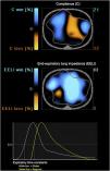
A 61-year-old patient with a history of COPD was in the postoperative phase of a right single lung transplant, requiring a high-flow oxygen cannula due to acute hypoxemic respiratory failure. An electrical impedance tomography (Pulmovista V500 - Dräger, Germany) was used to assess regional changes in tidal ventilation and end-expiratory lung volume (EELV). An inspiratory flow rate of 60L/min and an inspired oxygen fraction of 0.4 were employed. Fig. 1 shows the changes in VT before and after the use of HFNC. In the non-transplanted lung, an increase in aeration is observed due to regional elevation of EELV (blue area) caused by increased expiratory resistance, leading to air trapping and alveolar overdistension (orange area). Consequently, expiratory time constants were prolonged (Fig. 2).

Figure 1
(0.25MB).
Figure 2
(0.29MB).
Financial support

This research did not receive any specific grant from funding agencies in the public, commercial, or not-for-profit sectors.

Conflicts of interest

None.

Copyright © 2023. Elsevier España, S.L.U. and SEMICYUC
Idiomas
Medicina Intensiva (English Edition)
Article options
Tools
es en

¿Es usted profesional sanitario apto para prescribir o dispensar medicamentos?

Are you a health professional able to prescribe or dispense drugs?