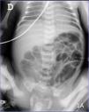
Premature 28-week baby weighing 850g at birth. Red blood cells are transfused due to prematurity anemia at the patient’s 22nd day of life. Twelve hours into the transfusion the patient shows abdominal distension and vomiting of gastric content. The blood test performed showed no alterations in the blood gasometry or elevated parameters of infection. The abdominal X-ray performed showed no signs suggestive of enterocolitis (Fig. 1A), but the abdominal ultrasound performed did show lack of peristalsis and intestinal loops with 2 layers of echogenic foci in the walls (white arrows), a sign indicative of the presence of intraluminal air (Fig. 2A), and echogenic foci in the hepatic parenchyma indicative of gas bubbles inside the portal veins (white arrows), a sign suggestive of portal venous gas (Fig. 2B) oriented as a clinical sign of enterocolitis. The patient remains on a diet and antibiotic therapy is started. The ultrasound looks normal with good disease progression (white asterisks) (Fig. 2C) after the completion of a 14 day course of antibiotic treatment.
Please cite this article as: Batista Muñoz A, Otero Vaccarello O, Rodriguez-Fanjul J. ¿Debería ser la ecografía abdominal la primera prueba de elección de imagen en la enterocolitis necrosante? Med Intensiva. 2021;45:445–446.


