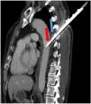
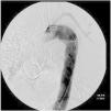
This is the case of a 54-year-old man treated with emergency vascular and thoracic surgery following a stab wound to the posterior thoracic wall at T4–T5 level. The patient’s hemodynamic and respiratory status is stable. Study was completed with a computed tomography (CT) scan. Both in the sagittal view (Fig. 1) and in the 3D reconstruction with software (Fig. 2-f, green arrow) the tip of the bladed weapon was found to be located at the descending aorta middle wall level surrounded by periaortic hematoma (Fig. 1, blue arrow) and an aortic mural hematoma (Fig. 1, red arrow). Then, a follow-up arteriography was performed at the operating room. Simultaneously, the knife was extracted and a thoracic stent-graft was deployed and later examined in the absence of bleeding (Fig. 3). Also, posterior selective embolization of the left subclavian artery distal branches was performed followed by hemostasis of paravertebral musculature. The patient’s clinical course at the intensive care unit (ICU) was favorable.
Please cite this article as: Barber Ansón M, Orera Pérez A, López Sala P. Laceración traumática de aorta torácica por herida de arma blanca. Med Intensiva. 2022;46:596–597.


