


To evaluate the diagnostic accuracy of diaphragmatic ultrasound in predicting extubation success.
DesignA diagnostic accuracy study was carried out.
ScopeIntensive Care Unit of an Academic hospital in the city of Bogotá (Colombia).
Patients or participantsA consecutive sample of patients >18 years of age subjected to invasive mechanical ventilation for >48h.
InterventionsDiaphragmatic ultrasound evaluation at the end of spontaneous ventilation testing.
Main variables of interestDiaphragmatic excursion (DE, cm), inspiration time (TPIAdia, s), diaphragm contraction speed (DE/TPIAdia, cm/s) and total time (Ttot, s) were evaluated, together with thickening fraction (TFdi, %).
ResultsA total of 84 patients were included, 79.8% (n=67) with successful extubation and 20.2% (n=17) with failed extubation. The variable with the best discriminatory capacity in predicting extubation success was diaphragm contraction speed, with AUC-ROC 0.70 (p=0.008).
ConclusionsDiaphragm contraction speed exhibited acceptable discriminatory capacity. Ultrasound could be part of a multifactorial approach in the extubation process.
Evaluar la exactitud diagnóstica de la ecografía diafragmática para predecir el éxito en la extubación.
DiseñoEstudio de exactitud diagnóstica.
ÁmbitoUnidad de Cuidado Intensivo Médico de un hospital académico de la ciudad de Bogotá (Colombia).
Pacientes o participantesMuestra consecutiva de pacientes mayores de 18 años con ventilación mecánica invasiva durante más de 48h.
IntervencionesEvaluación ecográfica diafragmática al finalizar la prueba de ventilación espontánea.
Variables de interés principalesSe evaluó la excursión diafragmática (ED, cm), el tiempo de inspiración (TPIAdia, s), la velocidad de contracción del diafragma (ED/TPIAdia, cm/s), el tiempo total (Ttot, s) y la fracción de engrosamiento (TFdi, %).
ResultadosSe incluyeron 84 pacientes, el 79.8% (n=67) con extubación exitosa y el 20,2% (n=17) con extubación fallida. La variable con mejor capacidad discriminatoria para predecir éxito en la extubación fue la velocidad de contracción, con un AUC-ROC de 0,70 (p=0,008).
ConclusionesLa velocidad de contracción diafragmática mostró una capacidad discriminatoria aceptable. La ultrasonografía podría formar parte de un abordaje multifactorial en el proceso de extubación.
The need for mechanical ventilation (MV) is one of the main reasons for admission to the Intensive Care Unit (ICU)1. Despite its benefits, however, the complications of MV are an important source of patient morbidity–mortality2–4. Establishing the optimum moment for withdrawal of ventilatory support remains one of the greatest challenges for the treating professional team, since late extubation is directly associated to an increased incidence of in-hospital infections, including ventilator-associated pneumonia (VAP), as well as to increased costs, diaphragm dysfunction, worsened quality of life over the middle term, and a longer stay in the ICU and in hospital in general5–7. In contrast, early extubation resulting in a need for reintubation has been associated to a 25–50% increase in patient mortality8,9.
The heterogeneity of the patients admitted to the ICU implies that the causes of extubation failure are also multiple10; diaphragm dysfunction appears to be implicated in up to 50% of all failed extubations11. This situation is related to the structural and functional changes observed in the muscle fibers after the start of ventilatory support12,13. On the basis of the above, one of the cornerstones of patient management is the facilitation of early rehabilitation1,14. To date, no reference parameters have been able to predict extubation success. The most widely used clinical parameters are the rapid shallow breathing index (RSBI), vital capacity (VC) and peak inspiratory pressure (PImax), among others15,16. There is great variability in the cut-off points and diagnostic precision of these parameters11, and none of them reflect the integrity of diaphragm structure and function.
In this context, in recent years ultrasound at the patient bedside (point of care) has become one of the tools of choice in the ICU due to its accessibility and low cost. It allows us to assess structure and function quantitatively and qualitatively before, during and after extubation17. A range of ultrasound parameters have been studied to date: diaphragmatic excursion (DE), thickening fraction (TFdi), contraction velocity (V)18–20 and even variations in rapid shallow breathing index (respiratory frequency/DE)21. The cut-off points of these parameters are likewise diverse, with great variability in performing the test.
The present study was carried out to evaluate the diagnostic accuracy of diaphragmatic ultrasound at the patient bedside in predicting extubation success.
Patients and methodsA prospective, observational cohort study on diagnostic accuracy was carried out.
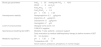
Study populationThe study was carried out in the ICU of an academic institution in the city of Bogotá (Colombia), with the consecutive inclusion of all patients over 18 years of age subjected to invasive MV for over 48h and who met the following inclusion criteria: (1) resolution of the cause of respiratory failure; (2) hemodynamic stability; (3) metabolic equilibrium; (4) optimum level of consciousness; and (5) indication of spontaneous breathing test (SBT) (Table 1).
Inclusion criteria.
| Blood gas parameters | PaO2≥55mmHg with FiO2<40% |
| PEEP<8cm H2O | |
| PaO2/FiO2>175 | |
| PaO2/PAO2>0.3 | |
| pH≥7.35≤7.48 | |
| Hemodynamic stability | Norepinephrine <0.1μg/kg/min |
| Dopamine <5μg/kg/min | |
| Dobutamine <5μg/kg/min | |
| Hemoglobin ≥7g/dl | |
| Level of consciousness | Glasgow coma score ≥12 |
| CAM-ICU negative | |
| Spontaneous breathing test (SBT) | Modality: T-tube: patients – pressure support |
| Daily evaluation by medical staff/respiratory therapy to define moment of SBT | |
| 30min duration | |
| Metabolic equilibrium | pH≥7.35≤7.48 |
| Temperature ≤38°C | |
| Serum sodium, potassium, phosphorus in normal ranges |
The exclusion criteria were: (1) neuromuscular disease; (2) previous diaphragmatic paralysis; (3) use of neuromuscular blockers during admission to the Unit; (4) pneumothorax or pneumomediastinum; and (5) pregnancy.
The following data were compiled at baseline: patient age and gender, cause of respiratory failure, duration of MV, and arterial gas and laboratory test values before extubation.
MeasurementsThe decision to perform the spontaneous breathing test (SBT) was assessed daily by the supervising medical team and the respiratory therapy group of the Unit. After the 30min of the SBT, diaphragmatic function was assessed by ultrasound, with calculation of the rapid shallow breathing index as part of the standard evaluation for establishing extubation.
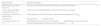
The diaphragmatic measurements were carried out by intensivists trained in ultrasound in the critical care setting, using a Sonocare ultrasound system (Sonosite EDGE 03VRYF). A 1–5MHz transducer was used for the M-mode evaluation of diaphragmatic excursion (DE, cm), time to peak inspiratory amplitude (TPIAdia, s), contraction velocity of the diaphragm (DE/TPIAdia, cm/s) and total time (Ttot, s). The thickening fraction (TFdi, %) was evaluated with a 6–13MHz transducer in M-mode (Fig. 1 and Table 2). Diaphragm dysfunction was defined as DE<1cm or paradoxical motion18.
![Ultrasound measures used to assess the success of extubation. Mean in M-mode. 1.1: Thickening fraction (TFdi, %), expiratory thickness (A), inspiratory thickness (B). 1.2: Measurement of diaphragmatic excursion (a) (DE, cm), time to peak inspiratory amplitude (b) (TPIAdia, s), diaphragmatic contraction velocity (DE/TPIAdia [cm/s]). 1.3: Time to peak inspiratory amplitude (a) (TPIAdia, s), total time (b) (s).](https://static.elsevier.es/multimedia/21735727/0000004500000004/v1_202104220744/S2173572720300795/v1_202104220744/en/main.assets/gr1.jpeg?xkr=dfhUZh+cpCdqta+flcPGXAWdthJkl6OkjGi2wxVdQHugE7U2BZXif5KTbqkTg4MratW5Cd8ifZQkDzESo7EccYU4UY/GvBjQBCVwjJ8fnmVZJspY4qSs/1koJScFavsrXJOpifyuAWdFlA3S08U270+x04gqGGOpbi0LyK31DSOJmD9eZmNCfckHwizpRCqWZ+FGdupnYF5f0UeOr3G2eO47rZVYPun5qeVnVWNRlkzstt2mBQhCB/IdPc0oaVVBhGcSZOrKvghNCq0M20qp1g== "Ultrasound measures used to assess the success of extubation. Mean in M-mode. 1.1: Thickening fraction (TFdi, %), expiratory thickness (A), inspiratory thickness (B). 1.2: Measurement of diaphragmatic excursion (a) (DE, cm), time to peak inspiratory amplitude (b) (TPIAdia, s), diaphragmatic contraction velocity (DE/TPIAdia [cm/s]). 1.3: Time to peak inspiratory amplitude (a) (TPIAdia, s), total time (b) (s).")
Ultrasound measures used to assess the success of extubation. Mean in M-mode. 1.1: Thickening fraction (TFdi, %), expiratory thickness (A), inspiratory thickness (B). 1.2: Measurement of diaphragmatic excursion (a) (DE, cm), time to peak inspiratory amplitude (b) (TPIAdia, s), diaphragmatic contraction velocity (DE/TPIAdia [cm/s]). 1.3: Time to peak inspiratory amplitude (a) (TPIAdia, s), total time (b) (s).
Diaphragmatic measurements.
| Measurement | Evaluation M-mode |
|---|---|
| Diaphragmatic excursion (DE, cm) | Excursion amplitude from start of contraction to maximum inspiration |
| Time to peak inspiratory amplitude (TPIAdia, s) | Time from start of diaphragmatic contraction to maximum inspiration |
| Diaphragmatic contraction velocity (cm/s) | Diaphragmatic excursion (DE)/time to peak inspiratory amplitude (TPIAdia) |
| Total time (s) | Inspiratory time+expiratory time |
| Thickening fraction (TFdi, %) | Diaphragmatic thickness at end of inspiration−diaphragmatic thickness at end of expiration/diaphragmatic thickness at end of expiration×100 |
The measurements were made only in the right half of the diaphragm, with the patient in the semi-sitting position (headrest raised 45 degrees). The transducer was positioned just below the ribcage, between the clavicular midline and the anterior axillary line. The ultrasound beam was directed cephalad, perpendicular to the posterior third of the diaphragm. Three operators performed the ultrasound explorations in the ICU, distributed as follows: 45 explorations made by an intensivist during the morning shift, and 20 explorations each performed by two intensivists in the afternoon. Before the study, a 12-h training session with an expert radiologist was held to ensure standardization of the ultrasound measurements.
Before extubation, all patients were reconnected to their previous ventilation mode during 1h22.
Study objectivesThe primary study objective was to determine the accuracy of diaphragmatic ultrasound as a predictor of the success of weaning from MV. Successful extubation was defined as the capacity to maintain spontaneous breathing for over 48h without ventilatory assistance after extubation. Failed extubation in turn was defined as the need for patient reintubation in under 48h9.
As secondary objective, we evaluated the differences in extubation success or failure in relation to the different demographic, clinical and ultrasound parameters, and diaphragm dysfunction (defined as DE<1cm or paradoxical motion)18.
Ethical aspectsThe study protocol was approved by the local Ethics Committee (Ref. no.: 205 of 2014). The study was considered to pose minimal risks for patients according to resolution 8430 of 1993 of the Colombian Ministry of Health. Informed consent was obtained from all the participants.
Statistical analysisConvenience non-probability sampling was performed, calculating a sample size of 84 patients based on an estimated prevalence of 20% for extubation failure, with a sensitivity of 90% and a specificity of 86%, a 95% confidence interval with an area under the receiver operating characteristic curve (AUC-ROC) of 0.15, an alpha error of 0.05, and a statistical power of 80%23,24.
Central tendency and dispersion measures were used for the quantitative variables, and frequencies and percentages for the qualitative variables.
The patients were divided into two groups according to the primary outcome (extubation success or failure). The chi-squared test was used for the bivariate comparison of categorical variables. The Student t-test in turn was used for the comparison of continuous variables exhibiting a normal distribution, while parameters with a non-normal distribution were contrasted by means of the Mann–Whitney U-test. The quantitative variables with a non-parametric distribution that included follow-up time were subjected to negative binomial regression or Poisson analysis, depending on whether the standard deviation (SD) was greater or smaller than the mean of such variables.
We calculated the operator characteristics of each of the ultrasound measures to predict extubation success or failure, and ROC curves were plotted to establish the diagnostic accuracy of each of the ultrasound parameters. The point of maximum discriminating capacity was selected, based on the Youden index.
The AUC-ROC was interpreted as follows25: =0.5, no discriminating capacity; >0.7–0.79: acceptable discriminating capacity; >0.8–0.89: excellent discriminating capacity; >0.9: outstanding discriminating capacity.
The data were analyzed using the SPSS version 20 statistical package and MedCalc version 19.
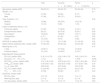
ResultsA total of 84 patients were included in the study, and no losses were recorded. The general characteristics of the study sample are described in Table 3. The median patient age was 58 years (range 35–51), with a female predominance (56%). Most of the patients (88%) presented medical conditions as the indication of MV, with an APACHE II severity score of 21 (17–28). The method of choice for SBT was the T-tube technique (85.7%), versus pressure support (14.3%).
General characteristics of the study sample. Total population, extubation success and failure groups.
| Total | Success | Failure | p | |
|---|---|---|---|---|
| n=67 (79.8%) | n=17 (20.2%) | |||
| Age (years), median (IQR) | 58 (35–51) | 58 (34–72) | 59 (49–64) | 0.79 |
| Gender, n (%) | 0.4 | |||
| Female | 47 (56) | 39 (83) | 8 (17) | |
| Male | 37 (44) | 28 (75.7) | 9 (24.3) | |
| Type of patient, n (%) | 0.39 | |||
| Medical | 74 (88) | 58 (78.4) | 16 (21.6) | |
| Surgical | 10 (12) | 9 (90) | 1 (10) | |
| Cause of respiratory failure, n (%) | ||||
| Pulmonary sepsis | 21 (25) | 17 (80.9) | 4 (19.1) | 0.87 |
| Extrapulmonary sepsis | 26 (31) | 20 (76.9) | 6 (23.1) | 0.56 |
| Neurological | 19 (22.6) | 15(78.9) | 4 (21.1) | 0.92 |
| Postoperative | 6 (7.1) | 6 (100) | 0 (0) | 0.20 |
| Others | 12 (14.3) | 9 (75) | 3 (25) | 0.65 |
| APACHE II, median (IQR) | 21 (17–28) | 19 (17–28) | 21 (18–25) | 0.52 |
| Rapid shallow breathing index, median (IQR) | 47 (36–63) | 48 (36–64) | 40 (32–62) | 0.38 |
| Weaning test, n (%) | 0.73 | |||
| T-tube | 72 (85.7) | 57 (79.2) | 15 (20.8) | |
| Pressure support | 12 (14.3) | 10 (83.3) | 2 (16.7) | |
| Diaphragmatic measurements | ||||
| DE (cm), median (IQR) | 2.18 (1.6–2.75) | 2.22 (1.66–2.75) | 2.02 (1.63–2.31) | 0.44 |
| Excursion <10mm, n (%) | 1 (1.2) | 0 (0) | 1 (100) | 0.046 |
| DE/TPIAdia (cm/s), median (IQR) | 2.74 (1.90–3.33) | 2.90 (2.00–4.01) | 2.02 (1.49–2.80) | 0.013 |
| TPIAdia (s), median (IQR) | 0.79 (0.64–1.02) | 0.80 (0.67–0.95) | 0.77 (0.62–1.08) | 0.76 |
| Ttot (s), mean (DE) | 2.96 (0.65) | 2.97 (0.65) | 2.93 (0.65) | 0.84 |
| TFdi (%), median (IQR) | 31 (24–45) | 32 (27–47) | 30 (21–35) | 0.10 |
| TFdi>30%, n (%) | 47 (56) | 39 (83) | 8 (17) | 0.40 |
| Outcomes, median (IQR) | ||||
| MV time, days | 5 (3–10) | 5 (3–9) | 7 (4–11) | 0.98 |
| Weaning time, days | 2 (1–3) | 2 (1–3) | 3 (1–3) | 0.65 |
| ICU stay (days), median (IQR) | 10 (7–17) | 10 (6–17) | 11 (8–16) | 0.69 |
APACHE II: Acute Physiology and Chronic Health Evaluation II; SD: standard deviation; DE: diaphragmatic excursion; IQR: interquartile range; TFdi: thickening fraction; TPIAdia: time to peak inspiratory amplitude; Ttot: total time; ICU: Intensive Care Unit; V: contraction velocity; MV: mechanical ventilation.
Successful extubation was achieved in 79.8% of the patients (n=67), and extubation failed in the remaining 20.2% of the cases (n=17). The comparison of results between both groups is shown in Table 3. There were no significant differences between the groups in terms of the demographic or clinical characteristics. However, the patients with failed extubation presented APACHE II scores that were slightly higher than those recorded in the patients with successful extubation. The rapid shallow breathing index was also similar in both groups, with slightly higher scores among the patients with successful extubation versus those with failed extubation: 48 (36–64) and 40 (32–62), respectively. The SBT choice likewise showed no significant differences. The duration of MV and of ICU stay was slightly longer in the failed extubation group, though the differences failed to reach statistical significance.
Of the different ultrasound parameters, differences were only observed for contraction velocity and diaphragm dysfunction (DE<1cm); the latter was only present in 1.2% of the total cases. The ROC curve for diaphragm contraction velocity is shown in Fig. 2. This variable presented AUC 0.70 (p=0.008; 95% CI: 0.58–0.79). Three cut-off points were calculated (shown in Table 4). The point of maximum discriminating capacity according to the Youden index was >2.9cm/s, with a sensitivity of 4.27% (95% CI: 0.34–0.59) and a specificity of 88.24% (95% CI: 0.50–0.93). Values of over 1.74cm/s were associated to greater sensitivity (85% [95% CI: 0.74–0.93]) and lesser specificity (41% [95% CI: 0.19–0.67]). On the other hand, thresholds of over 4.3cm/s showed a sensitivity of 19.4% (95% CI: 0.11–0.30) and a specificity of 88.24% (95% CI: 0.64–0.99).
![Receiver operating characteristic curve for contraction velocity and extubation success. AUC 0.70 (p=0.008 [95% CI: 0.58–0.79]).](https://static.elsevier.es/multimedia/21735727/0000004500000004/v1_202104220744/S2173572720300795/v1_202104220744/en/main.assets/gr2.jpeg?xkr=dfhUZh+cpCdqta+flcPGXAWdthJkl6OkjGi2wxVdQHugE7U2BZXif5KTbqkTg4MratW5Cd8ifZQkDzESo7EccYU4UY/GvBjQBCVwjJ8fnmVZJspY4qSs/1koJScFavsrXJOpifyuAWdFlA3S08U270+x04gqGGOpbi0LyK31DSOJmD9eZmNCfckHwizpRCqWZ+FGdupnYF5f0UeOr3G2eO47rZVYPun5qeVnVWNRlkzstt2mBQhCB/IdPc0oaVVBhGcSZOrKvghNCq0M20qp1g== "Receiver operating characteristic curve for contraction velocity and extubation success. AUC 0.70 (p=0.008 [95% CI: 0.58–0.79]).")
Operator characteristics.
| Contraction velocity (cm/s) | Sensitivity | Specificity | PPV | NPV | LR+ | LR− |
|---|---|---|---|---|---|---|
| >1.74 | 85 | 42 | 85 | 41 | 1.45 | 0.36 |
| >2.90 | 46.27 | 88.24 | 94 | 29 | 3.93 | 0.61 |
| >4.3 | 19.4 | 88.24 | 87 | 22 | 1.62 | 0.91 |
LR+: positive likelihood ratio; LR−: negative likelihood ratio; NPV: negative predictive value; PPV: positive predictive value.
The main finding of our investigation was an acceptable discriminating capacity (AUC 0.70; p=0.008 [95% CI: 0.58–0.79]) in predicting extubation success or failure on assessing diaphragmatic contraction velocity as isolated marker in critical patients admitted to the ICU.
A range of ultrasound parameters have been evaluated in the MV weaning process26. The most widely studied are DE and TFdi. Two meta-analyses have compiled the available evidence. The first included 19 observational studies with a total of 1068 patients. The DE values were between 10 and 27mm and the TFdi values between 20 and 36%. The analysis of the ROC curve for TFdi yielded AUC 0.87, while in the case of DE the lack of data and their heterogeneity only allowed the estimation of a cumulative specificity of 75% and a sensitivity of 80%27. The second meta-analysis, published in 2018, evaluated 13 observational studies with a total of 742 patients. The findings were similar to those of the previous meta-analysis, with good performance being observed for both DE and TFdi, with AUC 0.859 and 0.838, respectively28. Nevertheless, both meta-analyses were characterized by heterogeneity in defining extubation failure, in the selection of patients, the indication of intubation, and the selection of cut-off points. In our study, neither DE nor TFdi showed differences between the extubation success and failure groups. The only variable exhibiting statistically significant differences was diaphragmatic contraction velocity. This variable is taken to be an indirect measure of diaphragm contraction strength. In healthy individuals, the normal value is estimated to be 1.3±0.4cm/s. The role of this variable has not been widely studied to date, though higher values appear to be related to an increased probability of successful extubation29. We recorded values of >2.9cm/s (2.00–4.01) in the successful extubation group versus >2.02cm/s in the failed extubation group (1.49–2.80) (p=0.013). A number of cut-off points were analyzed with a view to optimizing the discriminating capacity of the test. A velocity >1.74cm/s showed high sensitivity. However, the low associated specificity could indicate a greater number of patients at risk of reintubation. On the other hand, thresholds above 4.3cm/s (sensitivity 19.4% and specificity 88.24%) would limit the start of weaning in clinical practice. The maximum discriminating capacity was established at >2.9cm/s, with only acceptable overall performance (AUC 0.70; p=0.008). To date, three studies have defined velocity as a differential marker in the extubation process. The first study described much lower thresholds than those established in our study (>0.8cm/s), with a sensitivity of 100%, a specificity of 86.67% and an outstanding discriminating capacity (AUC 0.93)29. The second study, with slightly higher thresholds (0.92cm/s), reported a sensitivity of 100%, with low specificity (45%) in discriminating extubation success (AUC 0.66)30. Lastly, the third study evaluated TPIAdia as derived variable (DE/TPIAdia), with a discriminating capacity similar to that found in our study (AUC 0.71)11.
The optimum time for suspending MV remains a challenge for multidisciplinary teams in the ICU. The need for reintubation is one of the most feared complications, because of the associated increase in patient mortality8. The incidence of extubation failure reported in the literature ranges from 10 to 25%31, which coincides with the figure recorded in our study (20.2%).
Muscle trauma with consequent ventilator-induced diaphragm dysfunction (VIDD) is one of the main causes of failed ventilation withdrawal32. In some cases it may go unnoticed; active search on the part of the clinician is therefore essential in this regard33. To date, the parameter used to evaluate diaphragm function has been direct fluoroscopy, though this technique is of limited applicability in the intensive care setting because of the problems posed by having to transfer critical patients outside the Unit11. For this reason diaphragmatic ultrasound has been found to be very useful as a tool that can be used at the patient bedside. In our study, diaphragm dysfunction was only observed in 1.2% of the cases, in contrast to other series in the literature that have reported a prevalence of 23–36%12. Ultrasound definition also has limitations, and the technique has not been prospectively contrasted versus the reference standards. Furthermore, the cut-off points described in the literature are heterogeneous. Nevertheless, we consider that the low frequency of diaphragm dysfunction recorded in our study is attributable to the presence of a strict early rehabilitation program that starts upon patient admission to the Unit.
Prolonged MV is one of the consequences of diaphragm dysfunction34. Our results showed a relatively short period of MV, with a median of 5 days, and with no differences between the extubation success and failure groups. Likewise, the ventilation weaning period corresponded to 40% of the ventilation time, in coincidence with the data found in the literature35. The total duration of stay in the Unit was slightly longer in the failure group, though statistical significance was not reached, probably because of statistical power limitations related to the sample size involved.
Taking into account that no clinical or imaging parameter considered isolatedly has been able to predict the outcome of the extubation process, the heterogeneity of the critically ill makes it necessary to adopt a multimodal approach to ventilation withdrawal36. The multiple systems involved should be evaluated qualitatively and quantitatively as part of the extubation process. Ultrasound could be a tool accompanying traditional evaluation, and in this sense the results of our study with regard to the rapid shallow breathing index (<105 in both groups) confirm its scant clinical usefulness when considered isolatedly. At present there are two main scenarios for ultrasound utilization at the critical patient bedside: (a) application as an evaluation and follow-up strategy in order to avoid muscle trauma32,37 and (b) use as part of multifunctional cardiovascular, pulmonary and pleural assessment38. We consider the definition of new integrating indices contemplating systematic ultrasound evaluation to be a priority with a view to improving the outcomes of the extubation process.
The main strength of our study is the identification of contraction velocity as an independent variable for estimating the success of extubation. Despite only acceptable performance in the ROC curves, it deserves becoming the focus of future research.
Our study has a number of limitations. Ultrasound is operator-dependent. Inter- and intra-observer variability in the ventilation weaning scenario has been evaluated only in relation to the measurement of the diaphragmatic thickening fraction39. Our measurements were made by different operators without conducting concordance studies between them; the results of the other ultrasound variables therefore require validation. On the other hand, the lack of distinction between the types of ventilation withdrawal (easy, difficult or prolonged9) could modify ultrasound performance as a prognostic tool. In turn, the severity of disease of the patients included in the study, assessed by means of the APACHE II score, was considered to be high; the findings therefore might not be extrapolatable to other ICUs of lesser complexity.
AuthorshipFabio Varon-Vega, Angela Hernandez, Luis Fernando Giraldo: study design and structure.
Mauricio Lopez and Edgar Caceres: data collection.
Fabio Varon-Vega, Ana Maria Uribe, Luis Fernando Giraldo and Stephanie Crevoisier: data analysis and drafting of the manuscript.
Conflicts of interestThe authors declare that they have no conflicts of interest.
Thanks are due to the medical and nursing and respiratory therapy staff of the medical ICU of the Fundación Neumológica Colombiana.
Please cite this article as: Varón-Vega F, Hernández Á, López M, Cáceres E, Giraldo-Cadavid LF, Uribe-Hernandez AM, et al. Utilidad de la ecografía diafragmática para predecir el éxito en la extubación. Med Intensiva. 2021;45:226–233.

