array:24 [
  "pii" => "S217357272100076X"
  "issn" => "21735727"
  "doi" => "10.1016/j.medine.2021.07.002"
  "estado" => "S300"
  "fechaPublicacion" => "2021-10-01"
  "aid" => "1477"
  "copyright" => "Elsevier España, S.L.U. and SEMICYUC"
  "copyrightAnyo" => "2020"
  "documento" => "article"
  "crossmark" => 1
  "subdocumento" => "crp"
  "cita" => "Med Intensiva. 2021;45:e11-e14"
  "abierto" => array:3 [
    "ES" => true
    "ES2" => true
    "LATM" => true
  ]
  "gratuito" => true
  "lecturas" => array:1 [
    "total" => 0
  ]
  "Traduccion" => array:1 [
    "es" => array:19 [
      "pii" => "S0210569120300917"
      "issn" => "02105691"
      "doi" => "10.1016/j.medin.2020.03.002"
      "estado" => "S300"
      "fechaPublicacion" => "2021-10-01"
      "aid" => "1477"
      "copyright" => "Elsevier España, S.L.U. y SEMICYUC"
      "documento" => "simple-article"
      "crossmark" => 1
      "subdocumento" => "crp"
      "cita" => "Med Intensiva. 2021;45:e11-e14"
      "abierto" => array:3 [
        "ES" => true
        "ES2" => true
        "LATM" => true
      ]
      "gratuito" => true
      "lecturas" => array:1 [
        "total" => 0
      ]
      "es" => array:11 [
        "idiomaDefecto" => true
        "cabecera" => "<span class="elsevierStyleTextfn">Carta cient&#237;fica</span>"
        "titulo" => "La ecograf&#237;a doppler color es un m&#233;todo preciso para evaluar la posici&#243;n de la sonda nasog&#225;strica en pacientes cr&#237;ticos"
        "tienePdf" => "es"
        "tieneTextoCompleto" => "es"
        "paginas" => array:1 [
          0 => array:2 [
            "paginaInicial" => "e11"
            "paginaFinal" => "e14"
          ]
        ]
        "titulosAlternativos" => array:1 [
          "en" => array:1 [
            "titulo" => "Color doppler ultrasound is a precise method to evaluate the position of the nasogastric tube in critical ill patients"
          ]
        ]
        "contieneTextoCompleto" => array:1 [
          "es" => true
        ]
        "contienePdf" => array:1 [
          "es" => true
        ]
        "resumenGrafico" => array:2 [
          "original" => 0
          "multimedia" => array:7 [
            "identificador" => "fig0010"
            "etiqueta" => "Figura 2"
            "tipo" => "MULTIMEDIAFIGURA"
            "mostrarFloat" => true
            "mostrarDisplay" => false
            "figura" => array:1 [
              0 => array:4 [
                "imagen" => "gr2.jpeg"
                "Alto" => 1680
                "Ancho" => 3000
                "Tamanyo" => 299998
              ]
            ]
            "descripcion" => array:1 [
              "es" => "<p id="spar0010" class="elsevierStyleSimplePara elsevierViewall">Flujograma que resume el proceso de selecci&#243;n de pacientes&#46; SNG&#58; sonda nasog&#225;strica&#59; UCI&#58; unidad de cuidados intensivos&#46;</p>"
            ]
          ]
        ]
        "autores" => array:1 [
          0 => array:2 [
            "autoresLista" => "N&#46;B&#46; Acosta Pedemonte, D&#46;H&#46; Bagilet, N&#46;S&#46; Rocchetti, G&#46;V&#46; Torresan, N&#46;A&#46; Rodr&#237;guez, C&#46;J&#46; Settecase"
            "autores" => array:6 [
              0 => array:2 [
                "nombre" => "N&#46;B&#46;"
                "apellidos" => "Acosta Pedemonte"
              ]
              1 => array:2 [
                "nombre" => "D&#46;H&#46;"
                "apellidos" => "Bagilet"
              ]
              2 => array:2 [
                "nombre" => "N&#46;S&#46;"
                "apellidos" => "Rocchetti"
              ]
              3 => array:2 [
                "nombre" => "G&#46;V&#46;"
                "apellidos" => "Torresan"
              ]
              4 => array:2 [
                "nombre" => "N&#46;A&#46;"
                "apellidos" => "Rodr&#237;guez"
              ]
              5 => array:2 [
                "nombre" => "C&#46;J&#46;"
                "apellidos" => "Settecase"
              ]
            ]
          ]
        ]
      ]
      "idiomaDefecto" => "es"
      "Traduccion" => array:1 [
        "en" => array:9 [
          "pii" => "S217357272100076X"
          "doi" => "10.1016/j.medine.2021.07.002"
          "estado" => "S300"
          "subdocumento" => ""
          "abierto" => array:3 [
            "ES" => true
            "ES2" => true
            "LATM" => true
          ]
          "gratuito" => true
          "lecturas" => array:1 [
            "total" => 0
          ]
          "idiomaDefecto" => "en"
          "EPUB" => "https://multimedia.elsevier.es/PublicationsMultimediaV1/item/epub/S217357272100076X?idApp=WMIE"
        ]
      ]
      "EPUB" => "https://multimedia.elsevier.es/PublicationsMultimediaV1/item/epub/S0210569120300917?idApp=WMIE"
      "url" => "/02105691/0000004500000007/v1_202109210634/S0210569120300917/v1_202109210634/es/main.assets"
    ]
  ]
  "itemSiguiente" => array:18 [
    "pii" => "S2173572721000771"
    "issn" => "21735727"
    "doi" => "10.1016/j.medine.2021.07.003"
    "estado" => "S300"
    "fechaPublicacion" => "2021-10-01"
    "aid" => "1446"
    "copyright" => "Elsevier Espa&#241;a&#44; S&#46;L&#46;U&#46; and SEMICYUC"
    "documento" => "article"
    "crossmark" => 1
    "subdocumento" => "crp"
    "cita" => "Med Intensiva. 2021;45:e15-e17"
    "abierto" => array:3 [
      "ES" => true
      "ES2" => true
      "LATM" => true
    ]
    "gratuito" => true
    "lecturas" => array:1 [
      "total" => 0
    ]
    "en" => array:11 [
      "idiomaDefecto" => true
      "cabecera" => "<span class="elsevierStyleTextfn">Scientific Letter</span>"
      "titulo" => "Double trigger and Pseudo-reverse-trigger&#63;"
      "tienePdf" => "en"
      "tieneTextoCompleto" => "en"
      "paginas" => array:1 [
        0 => array:2 [
          "paginaInicial" => "e15"
          "paginaFinal" => "e17"
        ]
      ]
      "titulosAlternativos" => array:1 [
        "es" => array:1 [
          "titulo" => "Doble trigger y &#191;falso&#63; reverse trigger"
        ]
      ]
      "contieneTextoCompleto" => array:1 [
        "en" => true
      ]
      "contienePdf" => array:1 [
        "en" => true
      ]
      "resumenGrafico" => array:2 [
        "original" => 0
        "multimedia" => array:8 [
          "identificador" => "fig0010"
          "etiqueta" => "Figure 2"
          "tipo" => "MULTIMEDIAFIGURA"
          "mostrarFloat" => true
          "mostrarDisplay" => false
          "figura" => array:1 [
            0 => array:4 [
              "imagen" => "gr2.jpeg"
              "Alto" => 1180
              "Ancho" => 2091
              "Tamanyo" => 598687
            ]
          ]
          "detalles" => array:1 [
            0 => array:3 [
              "identificador" => "at0055"
              "detalle" => "Figure "
              "rol" => "short"
            ]
          ]
          "descripcion" => array:1 [
            "en" => "<p id="spar0010" class="elsevierStyleSimplePara elsevierViewall">Registry &#40;flow and volume&#41; of Paw&#44; Pes&#44; and Pves signs during pressure support ventilation in case &#35;2&#46; In this case&#44; we presented ventilation with double cycling defined as DTA due to auto-triggering of the ventilator preceding the patient&#8217;s respiratory driver &#40;reverse triggering in a general sense&#41;&#46; In this case&#44; the patient&#8217;s cycle does not start by contracting the inspiratory muscles&#44; but by releasing intense active expiration&#44; as confirmed by vesical pressure&#46; The contraction of the diaphragm comes after active exhalation release&#46; To think that this respiratory driver that includes forced exhalation release and the patient&#8217;s inspiratory effort is the product of some reflex does not seem very likely&#46; In this case the correlation between the mechanical and the neural cycles is variable&#46; The parameters associated with inspiratory effort are&#58; DPes&#58; 11&#46;55<span class="elsevierStyleHsp" style=""></span>&#177;<span class="elsevierStyleHsp" style=""></span>1&#46;22<span class="elsevierStyleHsp" style=""></span>cmH<span class="elsevierStyleInf">2</span>O&#44; note the higher DPes in double triggering&#59; WOB&#44; 1&#46;06<span class="elsevierStyleHsp" style=""></span>&#177;<span class="elsevierStyleHsp" style=""></span>0&#46;42<span class="elsevierStyleHsp" style=""></span>J&#47;L&#59; PTP&#44; 183&#46;95<span class="elsevierStyleHsp" style=""></span>&#177;<span class="elsevierStyleHsp" style=""></span>27&#46;59<span class="elsevierStyleHsp" style=""></span>cmH<span class="elsevierStyleInf">2</span>O<span class="elsevierStyleHsp" style=""></span>s&#47;min&#59; in this case&#44; work of breathing was associated with the volume generated by cycle stacking corresponding to double cycling&#46; Also&#44; the appearance of double cycling varied&#46;</p>"
          ]
        ]
      ]
      "autores" => array:1 [
        0 => array:2 [
          "autoresLista" => "J&#46;A&#46; Ben&#237;tez Lozano, C&#46; de la Fuente Martos, J&#46;M&#46; Serrano Sim&#243;n"
          "autores" => array:3 [
            0 => array:2 [
              "nombre" => "J&#46;A&#46;"
              "apellidos" => "Ben&#237;tez Lozano"
            ]
            1 => array:2 [
              "nombre" => "C&#46;"
              "apellidos" => "de la Fuente Martos"
            ]
            2 => array:2 [
              "nombre" => "J&#46;M&#46;"
              "apellidos" => "Serrano Sim&#243;n"
            ]
          ]
        ]
      ]
    ]
    "idiomaDefecto" => "en"
    "EPUB" => "https://multimedia.elsevier.es/PublicationsMultimediaV1/item/epub/S2173572721000771?idApp=WMIE"
    "url" => "/21735727/0000004500000007/v1_202109230629/S2173572721000771/v1_202109230629/en/main.assets"
  ]
  "itemAnterior" => array:18 [
    "pii" => "S2173572721001120"
    "issn" => "21735727"
    "doi" => "10.1016/j.medine.2020.03.006"
    "estado" => "S300"
    "fechaPublicacion" => "2021-10-01"
    "aid" => "1491"
    "copyright" => "Elsevier Espa&#241;a&#44; S&#46;L&#46;U&#46; and SEMICYUC"
    "documento" => "simple-article"
    "crossmark" => 1
    "subdocumento" => "crp"
    "cita" => "Med Intensiva. 2021;45:e7-e10"
    "abierto" => array:3 [
      "ES" => true
      "ES2" => true
      "LATM" => true
    ]
    "gratuito" => true
    "lecturas" => array:1 [
      "total" => 0
    ]
    "en" => array:10 [
      "idiomaDefecto" => true
      "cabecera" => "<span class="elsevierStyleTextfn">Scientific Letter</span>"
      "titulo" => "The first year of experience with an extracorporeal resuscitation program for refractory in-hospital cardiac arrest"
      "tienePdf" => "en"
      "tieneTextoCompleto" => "en"
      "paginas" => array:1 [
        0 => array:2 [
          "paginaInicial" => "e7"
          "paginaFinal" => "e10"
        ]
      ]
      "titulosAlternativos" => array:1 [
        "es" => array:1 [
          "titulo" => "Resucitacion extracorporea en la parada cardiorrespiratoria intrahospitalaria refractaria&#46; experiencia del primer a&#241;o de un programa de ECMO-RCP"
        ]
      ]
      "contieneTextoCompleto" => array:1 [
        "en" => true
      ]
      "contienePdf" => array:1 [
        "en" => true
      ]
      "autores" => array:1 [
        0 => array:2 [
          "autoresLista" => "J&#46;I&#46; Chico-Carballas, A&#46; Touceda-Bravo, S&#46; Freita-Ramos, D&#46; Mosquera-Rodriguez, V&#46; G&#243;mez-Casal, M&#46; Pi&#241;on-Esteban"
          "autores" => array:6 [
            0 => array:2 [
              "nombre" => "J&#46;I&#46;"
              "apellidos" => "Chico-Carballas"
            ]
            1 => array:2 [
              "nombre" => "A&#46;"
              "apellidos" => "Touceda-Bravo"
            ]
            2 => array:2 [
              "nombre" => "S&#46;"
              "apellidos" => "Freita-Ramos"
            ]
            3 => array:2 [
              "nombre" => "D&#46;"
              "apellidos" => "Mosquera-Rodriguez"
            ]
            4 => array:2 [
              "nombre" => "V&#46;"
              "apellidos" => "G&#243;mez-Casal"
            ]
            5 => array:2 [
              "nombre" => "M&#46;"
              "apellidos" => "Pi&#241;on-Esteban"
            ]
          ]
        ]
      ]
    ]
    "idiomaDefecto" => "en"
    "EPUB" => "https://multimedia.elsevier.es/PublicationsMultimediaV1/item/epub/S2173572721001120?idApp=WMIE"
    "url" => "/21735727/0000004500000007/v1_202109230629/S2173572721001120/v1_202109230629/en/main.assets"
  ]
  "en" => array:18 [
    "idiomaDefecto" => true
    "cabecera" => "<span class="elsevierStyleTextfn">Scientific Letter</span>"
    "titulo" => "Color doppler ultrasound is a precise method to evaluate the position of the nasogastric tube in critical ill patients"
    "tieneTextoCompleto" => true
    "saludo" => "Dear Editor&#58;"
    "paginas" => array:1 [
      0 => array:2 [
        "paginaInicial" => "e11"
        "paginaFinal" => "e14"
      ]
    ]
    "autores" => array:1 [
      0 => array:4 [
        "autoresLista" => "N&#46;B&#46; Acosta Pedemonte, D&#46;H&#46; Bagilet, N&#46;S&#46; Rocchetti, G&#46;V&#46; Torresan, N&#46;A&#46; Rodr&#237;guez, C&#46;J&#46; Settecase"
        "autores" => array:6 [
          0 => array:4 [
            "nombre" => "N&#46;B&#46;"
            "apellidos" => "Acosta Pedemonte"
            "email" => array:1 [
              0 => "Noelia_acosta_fcm@hotmail.com"
            ]
            "referencia" => array:1 [
              0 => array:2 [
                "etiqueta" => "&#42;"
                "identificador" => "cor0005"
              ]
            ]
          ]
          1 => array:2 [
            "nombre" => "D&#46;H&#46;"
            "apellidos" => "Bagilet"
          ]
          2 => array:2 [
            "nombre" => "N&#46;S&#46;"
            "apellidos" => "Rocchetti"
          ]
          3 => array:2 [
            "nombre" => "G&#46;V&#46;"
            "apellidos" => "Torresan"
          ]
          4 => array:2 [
            "nombre" => "N&#46;A&#46;"
            "apellidos" => "Rodr&#237;guez"
          ]
          5 => array:2 [
            "nombre" => "C&#46;J&#46;"
            "apellidos" => "Settecase"
          ]
        ]
        "afiliaciones" => array:1 [
          0 => array:2 [
            "entidad" => "Unidad de Cuidados Intensivos&#44; Hospital Escuela Eva Per&#243;n&#44; Granadero Baigorria&#44; Santa Fe&#44; Argentina"
            "identificador" => "aff0005"
          ]
        ]
        "correspondencia" => array:1 [
          0 => array:3 [
            "identificador" => "cor0005"
            "etiqueta" => "&#8270;"
            "correspondencia" => "Corresponding author&#46;"
          ]
        ]
      ]
    ]
    "titulosAlternativos" => array:1 [
      "es" => array:1 [
        "titulo" => "La ecograf&#237;a doppler color es un m&#233;todo preciso para evaluar la posici&#243;n de la sonda nasog&#225;strica en pacientes cr&#237;ticos"
      ]
    ]
    "resumenGrafico" => array:2 [
      "original" => 0
      "multimedia" => array:8 [
        "identificador" => "fig0010"
        "etiqueta" => "Figure 2"
        "tipo" => "MULTIMEDIAFIGURA"
        "mostrarFloat" => true
        "mostrarDisplay" => false
        "figura" => array:1 [
          0 => array:4 [
            "imagen" => "gr2.jpeg"
            "Alto" => 1680
            "Ancho" => 3000
            "Tamanyo" => 299998
          ]
        ]
        "detalles" => array:1 [
          0 => array:3 [
            "identificador" => "at0035"
            "detalle" => "Figure "
            "rol" => "short"
          ]
        ]
        "descripcion" => array:1 [
          "en" => "<p id="spar0010" class="elsevierStyleSimplePara elsevierViewall">Flowchart including the patient selection process&#46; ICU&#44; intensive care unit&#59; NGT&#44; nasogastric tube&#46;</p>"
        ]
      ]
    ]
    "textoCompleto" => "<span class="elsevierStyleSections"><p id="par0005" class="elsevierStylePara elsevierViewall">Early enteral nutrition in critically ill patients is essential to preserve the function of villi in the small intestine&#44; regulate immunity&#44; and the systemic inflammatory response&#46;<a class="elsevierStyleCrossRef" href="#bib0005"><span class="elsevierStyleSup">1</span></a> Enteral nutrition support is often performed through tubes&#44; being the nasogastric tube &#40;NGT&#41; the one most commonly used&#46;<a class="elsevierStyleCrossRef" href="#bib0010"><span class="elsevierStyleSup">2</span></a> Although the rate of complications associated with the insertion of NGTs is very low&#44; some of them can be serious&#44; especially with the NGT has been inserted in an anomalous position &#40;like the airway&#41;&#46;<a class="elsevierStyleCrossRefs" href="#bib0015"><span class="elsevierStyleSup">3&#8211;5</span></a> For this reason&#44; the insertion of the NGT should be performed by trained personnel&#46; Also&#44; it is mandatory to confirm the correct position of the NGT using different techniques&#46; The techniques available today are based on tests like the &#171;bubbling&#187; testing &#40;auscultation of bubble sounds following the entry of air&#41;&#44; pH stripes to test acidity or the interpretation of ultrasound images&#46; However&#44; the fact that these tests are rather unreliable added to the cost and poor interpretation of the last 2 make it necessary to have another low-cost and useful method at the patient&#8217;s bedside like the ultrasound that is widely available in the intensive care units &#40;ICU&#41; setting&#46;</p><p id="par0010" class="elsevierStylePara elsevierViewall">The objective of this study was to assess the diagnostic capabilities of the color Doppler ultrasound &#40;CDU&#41; to detect the position of the NGT in adult critically ill patients&#46;</p><p id="par0015" class="elsevierStylePara elsevierViewall">Back in 2019&#44; a prospective and observational study was conducted at a high-complexity ICU for 4 months&#46; This study was approved by the local ethics committee&#46; Prior to their inclusion in the study&#44; the patients or their families gave their written informed consent&#46;</p><p id="par0020" class="elsevierStylePara elsevierViewall">Patients over 18 with an indication for NGT insertion for total or supplementary enteral nutrition at the ICU setting were included</p><p id="par0025" class="elsevierStylePara elsevierViewall">The following patients were excluded&#58; <span class="elsevierStyleItalic">a&#41;</span> patients with congenital or acquired anatomical disorders that would complicate the insertion of the NGT&#44; <span class="elsevierStyleItalic">b&#41;</span> epistaxis within the previous 7 days&#44; <span class="elsevierStyleItalic">c&#41;</span> facial trauma or basilar skull fracture&#44; <span class="elsevierStyleItalic">d&#41;</span> persistent vomiting&#44; <span class="elsevierStyleItalic">e&#41;</span> suspected or confirmed esophageal perforation&#44; <span class="elsevierStyleItalic">f&#41;</span> severe coagulation disorder&#44; <span class="elsevierStyleItalic">g&#41;</span> active digestive bleeding&#44; and <span class="elsevierStyleItalic">h&#41;</span> previous digestive tract surgery or immediate postoperative&#46;</p><p id="par0030" class="elsevierStylePara elsevierViewall">The most commonly used tube for enteral nutrition is the 125 cm long&#44; 10-Fr caliber Silmag&#174; gastrostomy tube &#40;with 3&#46;3 mm of external diameter&#41;&#46; This NGT is a tungsten-weighted tube with a semi-rigid guidewire&#46; Experienced medical personnel inserted it and after the instrumentation the stomach was observed to confirm the position of the NGT&#46; The Mindray3C5P &#40;2&#8211;5 MHz&#41; convex ultrasound transducer &#40;China&#41; was used&#46; Using an epigastric window&#44; the gastric antrum was identified in bidimensional mode&#46; After visualization of the gastric antrum&#44; a bolus of 20 mL of air was injected through the NGT&#46; At the same time&#44; both in the epigastric window and in the duplex ultrasound &#40;simultaneous two-dimensional mode and color Doppler ultrasound&#41; the presence of a jet or the sudden appearance of a Doppler sign was confirmed &#40;<a class="elsevierStyleCrossRef" href="#fig0005">Fig&#46; 1</a> and video of the Supplementary data&#41;&#46; The ultrasounds were performed by trained intensivists in the use of this method&#46; The position of the NGT was considered correct when air was instilled&#44; and the jet was evident in the stomach&#46; On the contrary&#44; the position of the NGT was considered incorrect&#46;</p><elsevierMultimedia ident="fig0005"></elsevierMultimedia><p id="par0035" class="elsevierStylePara elsevierViewall">Immediately after performing the ultrasound&#44; an X-ray was used to confirm and monitor the correct placement of the NGT that was interpreted by an expert radiologist in diagnostic imaging modalities and who had been blind to the NGT insertion and the ultrasound&#46; The placement of the NGT was considered correct only through this method when the NGT distal border was found in the stomach&#46;</p><p id="par0040" class="elsevierStylePara elsevierViewall">Variables registered at admission&#58; sex&#44; age&#59; Acute Physiology and Chronic Health Evaluation II &#40;APACHE II&#41;&#44; Simplified Acute Physiology Score II &#40;SAPS II&#41;&#44; and disease &#40;clinical&#44; trauma&#44; surgical&#41;&#46;</p><p id="par0045" class="elsevierStylePara elsevierViewall">Variables selected on the day of NGT insertion&#58; Sequential Organ Failure Assessment score &#40;SOFA&#41;&#59; mechanical ventilation &#40;MV&#41;&#59; tracheostomy&#59; indication for NGT &#40;oral-enteral nutrition&#44; enteral nutrition&#44; enteral-parenteral nutrition&#41;&#59; days of ICU stay&#59; ultrasound diagnosis of the position of the NGT&#59; X-ray monitorization&#44; and time until the diagnosis of each imaging modality&#46;</p><p id="par0050" class="elsevierStylePara elsevierViewall">A descriptive analysis of the qualitative variables was performed&#46; These variables were expressed as frequencies and percentages&#46; Quantitative variables were expressed as mean and standard deviation or&#44; for asymmetric distributions&#44; as median and interquartile range &#40;p25&#8211;p75&#41;&#46; The findings seen on the X-ray were considered the gold standard and used to estimate sensitivity&#44; the positive predictive values &#40;PPV&#41;&#44; and the negative predictive values &#40;NPV&#41;&#46; The association between the variables &#40;ultrasound diagnosis of the position of the NGT&#44; and X-ray monitorization&#41; was established using Spearman&#8217;s correlation coefficient&#46; The Wilcoxon test was used to compare the necessary time to achieve the diagnosis of the position of the NGT using the 2 methods&#46;</p><p id="par0055" class="elsevierStylePara elsevierViewall"><span class="elsevierStyleItalic">P</span> values &#60; &#46;05 were considered statistically significant&#46; The statistical analysis was performed using the statistical software SPSS&#174; v&#46;22&#46;0&#46;</p><p id="par0060" class="elsevierStylePara elsevierViewall">During the 4 months&#44; a total of 196 patients were admitted to the ICU&#44; 88 of whom required NGT insertion&#46; A total of 17 of these patients were excluded because the NGT requirement was different from nutrition &#40;<a class="elsevierStyleCrossRef" href="#fig0010">Fig&#46; 2</a>&#41;&#46;</p><elsevierMultimedia ident="fig0010"></elsevierMultimedia><p id="par0065" class="elsevierStylePara elsevierViewall">The demographic variables&#44; APACHE II&#44; SAPS II&#44; and disease at ICU admission are shown on <a class="elsevierStyleCrossRef" href="#tbl0005">Table 1</a>&#46; The details of the variables registered at the moment when the NGT was inserted are shown on <a class="elsevierStyleCrossRef" href="#tbl0010">Table 2</a>&#46;</p><elsevierMultimedia ident="tbl0005"></elsevierMultimedia><elsevierMultimedia ident="tbl0010"></elsevierMultimedia><p id="par0070" class="elsevierStylePara elsevierViewall">The position of the NGT was correct according to the CDU 32 times and according to the X-ray 31 times&#46; In ambiguous cases&#44; the correct placement of the NGT was identified in the lower third of the esophagus through X-ray&#46; The position was considered incorrect according to both imaging modalities on 5 different occasions&#46;</p><p id="par0075" class="elsevierStylePara elsevierViewall">In this study&#44; the sensitivity&#44; PPV&#44; and NPV estimated to achieve diagnosis on the CDU were 100&#37;&#44; 96&#46;9&#37;&#44; and 100&#37;&#44; respectively&#46; Also&#44; a good correlation was found between the CDU and the X-ray &#40;Spearman&#8217;s correlation coefficient&#44; Rho&#44; 0&#46;898&#59; <span class="elsevierStyleItalic">P</span> &#60; &#46;001&#41;&#46;</p><p id="par0080" class="elsevierStylePara elsevierViewall">With respect to the necessary time to achieve diagnosis of the correct position of the NGT&#44; the median was 10 min &#40;IQR&#44; 9&#8211;15&#41; for the DCU&#44; and 90 min &#40;IQR&#44; 30&#8211;120&#41; for the X-ray &#40;<span class="elsevierStyleItalic">P &#60;</span> &#46;001&#41;&#46;</p><p id="par0085" class="elsevierStylePara elsevierViewall">Other authors have used ultrasound techniques to identify the NGT inside the gastric antrum with good sensitivity&#46;<a class="elsevierStyleCrossRefs" href="#bib0030"><span class="elsevierStyleSup">6&#8211;9</span></a> Kim et al&#46; used the two-dimensional ultrasound to guide the placement of the NGT and see it run down the cervical esophagus finding a 93&#37; sensitivity and a 98&#37; PPV&#46;<a class="elsevierStyleCrossRef" href="#bib0050"><span class="elsevierStyleSup">10</span></a> In approximately half of the cases&#44; additional maneuvers were required to make the imaging modality more sensitive&#44; and a high-resolution ultrasound probe was used for a better visualization of the esophagus&#44; but this is something that may not be reproducible in ICUs with fewer resources&#46;</p><p id="par0090" class="elsevierStylePara elsevierViewall">Zatelli and Vezzali studied ultrasound images in multiple windows &#40;including the instillation of fluid&#41; after NGT insertion without CDU guidance&#44; and they reported an 86&#46;4&#37; sensitivity&#44; an 66&#44;7&#37; specificity&#44; and a 97&#37; PPV&#46;<a class="elsevierStyleCrossRef" href="#bib0030"><span class="elsevierStyleSup">6</span></a> Our study found higher sensitivity and specificity rates and a similar predictive value despite the lower number of windows&#46; The advantage here is that the time required is shorter&#44; and that the presence of the doctor is not mandatory during NGT insertion&#44; meaning that it can be inserted by another healthcare professional for later checking&#46; In contrast with the aforementioned study&#44; we chose the instillation of air instead of saline solution due to the possibility of iatrogenesis due to an incorrect NGT insertion in the respiratory tract&#46;</p><p id="par0095" class="elsevierStylePara elsevierViewall">As far as we know&#44; there are no studies on the use of CDU for this purpose&#46; These are some of the limitations of our study we should mention&#58; the small sample size&#44; the populations that were not studied&#58; obesity&#44; pregnancy&#44; hiatal hernia&#44; GI surgery postoperative &#40;because no cases in our sample were found&#41;&#44; the use of a single ultrasound window&#44; and the indirect visualization of the nasogastric tube&#46; After the results obtained future studies should be conducted&#46; They should compare the CDU to the two-dimensional mode&#59; sensitivity and specificity changes to the combination of other ultrasound windows&#59; and secondary variations to different velocities during bolus administration&#46;</p><p id="par0100" class="elsevierStylePara elsevierViewall">The CDU proved to be a fast&#44; practical&#44; cost-effective&#44; and highly accurate imaging modality to check the position of the NGT in a small group of adult critically ill patients&#46; Although the CDU may be considered the first-line imaging modality for guidance purposes&#44; studies with larger populations are still needed to confirm these findings&#46;</p><span id="sec0005" class="elsevierStyleSection elsevierViewall"><span class="elsevierStyleSectionTitle" id="sect0005">Funding</span><p id="par0105" class="elsevierStylePara elsevierViewall">This study received no funding whatsoever&#46;</p></span><span id="sec0010" class="elsevierStyleSection elsevierViewall"><span class="elsevierStyleSectionTitle" id="sect0010">Conflicts of interest</span><p id="par0110" class="elsevierStylePara elsevierViewall">None reported&#46;</p></span></span>"
    "textoCompletoSecciones" => array:1 [
      "secciones" => array:4 [
        0 => array:2 [
          "identificador" => "sec0005"
          "titulo" => "Funding"
        ]
        1 => array:2 [
          "identificador" => "sec0010"
          "titulo" => "Conflicts of interest"
        ]
        2 => array:2 [
          "identificador" => "xack556903"
          "titulo" => "Acknowledgements"
        ]
        3 => array:1 [
          "titulo" => "References"
        ]
      ]
    ]
    "pdfFichero" => "main.pdf"
    "tienePdf" => true
    "NotaPie" => array:1 [
      0 => array:2 [
        "etiqueta" => "&#9734;"
        "nota" => "<p class="elsevierStyleNotepara" id="npar0005">Please cite this article as&#58; Acosta Pedemonte NB&#44; Bagilet DH&#44; Rocchetti NS&#44; Torresan GV&#44; Rodr&#237;guez NA&#44; Settecase CJ&#46; La ecograf&#237;a doppler color es un m&#233;todo preciso para evaluar la posici&#243;n de la sonda nasog&#225;strica en pacientes cr&#237;ticos&#46; Med Intensiva&#46; 2021&#59;45&#58;e11&#8211;e14&#46;</p>"
      ]
    ]
    "apendice" => array:1 [
      0 => array:1 [
        "seccion" => array:1 [
          0 => array:4 [
            "apendice" => "<p id="par0125" class="elsevierStylePara elsevierViewall">The following is Supplementary data to this article&#58;<elsevierMultimedia ident="upi0005"></elsevierMultimedia></p>"
            "etiqueta" => "Appendix A"
            "titulo" => "Supplementary data"
            "identificador" => "sec0020"
          ]
        ]
      ]
    ]
    "multimedia" => array:5 [
      0 => array:8 [
        "identificador" => "fig0005"
        "etiqueta" => "Figure 1"
        "tipo" => "MULTIMEDIAFIGURA"
        "mostrarFloat" => true
        "mostrarDisplay" => false
        "figura" => array:1 [
          0 => array:4 [
            "imagen" => "gr1.jpeg"
            "Alto" => 679
            "Ancho" => 1600
            "Tamanyo" => 97403
          ]
        ]
        "detalles" => array:1 [
          0 => array:3 [
            "identificador" => "at0030"
            "detalle" => "Figure "
            "rol" => "short"
          ]
        ]
        "descripcion" => array:1 [
          "en" => "<p id="spar0005" class="elsevierStyleSimplePara elsevierViewall">Comparison of images obtained in Doppler US mode prior to the instillation of air through nasogastric tube &#40;left&#41;&#44; and during the procedure &#40;right&#41;&#46;</p>"
        ]
      ]
      1 => array:8 [
        "identificador" => "fig0010"
        "etiqueta" => "Figure 2"
        "tipo" => "MULTIMEDIAFIGURA"
        "mostrarFloat" => true
        "mostrarDisplay" => false
        "figura" => array:1 [
          0 => array:4 [
            "imagen" => "gr2.jpeg"
            "Alto" => 1680
            "Ancho" => 3000
            "Tamanyo" => 299998
          ]
        ]
        "detalles" => array:1 [
          0 => array:3 [
            "identificador" => "at0035"
            "detalle" => "Figure "
            "rol" => "short"
          ]
        ]
        "descripcion" => array:1 [
          "en" => "<p id="spar0010" class="elsevierStyleSimplePara elsevierViewall">Flowchart including the patient selection process&#46; ICU&#44; intensive care unit&#59; NGT&#44; nasogastric tube&#46;</p>"
        ]
      ]
      2 => array:8 [
        "identificador" => "tbl0005"
        "etiqueta" => "Table 1"
        "tipo" => "MULTIMEDIATABLA"
        "mostrarFloat" => true
        "mostrarDisplay" => false
        "detalles" => array:1 [
          0 => array:3 [
            "identificador" => "at0040"
            "detalle" => "Table "
            "rol" => "short"
          ]
        ]
        "tabla" => array:2 [
          "leyenda" => "<p id="spar0020" class="elsevierStyleSimplePara elsevierViewall">APACHE II&#44; Acute Physiology and Chronic Health Evaluation II&#59; F&#44; female&#59; ICU&#44; intensive care unit&#59; M&#44; male&#59; SAPS II&#44; Simplified Acute Physiology Score II&#59; SD&#44; standard deviation&#46;</p>"
          "tablatextoimagen" => array:1 [
            0 => array:2 [
              "tabla" => array:1 [
                0 => """
                  <table border="0" frame="\n
                  \t\t\t\t\tvoid\n
                  \t\t\t\t" class=""><thead title="thead"><tr title="table-row"><th class="td" title="\n
                  \t\t\t\t\ttable-head\n
                  \t\t\t\t  " align="left" valign="\n
                  \t\t\t\t\ttop\n
                  \t\t\t\t" scope="col" style="border-bottom: 2px solid black">Variables&nbsp;\t\t\t\t\t\t\n
                  \t\t\t\t\t\t</th><th class="td" title="\n
                  \t\t\t\t\ttable-head\n
                  \t\t\t\t  " align="left" valign="\n
                  \t\t\t\t\ttop\n
                  \t\t\t\t" scope="col" style="border-bottom: 2px solid black">30 patients&nbsp;\t\t\t\t\t\t\n
                  \t\t\t\t\t\t</th></tr></thead><tbody title="tbody"><tr title="table-row"><td class="td-with-role" title="\n
                  \t\t\t\t\ttable-entry\n
                  \t\t\t\t ; entry_with_role_rowhead " align="left" valign="\n
                  \t\t\t\t\ttop\n
                  \t\t\t\t"><span class="elsevierStyleItalic">Age&#44; mean &#40;&#177; SD&#41;</span>&nbsp;\t\t\t\t\t\t\n
                  \t\t\t\t</td><td class="td" title="\n
                  \t\t\t\t\ttable-entry\n
                  \t\t\t\t  " align="left" valign="\n
                  \t\t\t\t\ttop\n
                  \t\t\t\t">46&#46;36 &#40;15&#46;88&#41;&nbsp;\t\t\t\t\t\t\n
                  \t\t\t\t</td></tr><tr title="table-row"><td class="td-with-role" title="\n
                  \t\t\t\t\ttable-entry\n
                  \t\t\t\t ; entry_with_role_rowhead " colspan="2" align="left" valign="\n
                  \t\t\t\t\ttop\n
                  \t\t\t\t"><span class="elsevierStyleItalic">Sex</span></td></tr><tr title="table-row"><td class="td-with-role" title="\n
                  \t\t\t\t\ttable-entry\n
                  \t\t\t\t ; entry_with_role_rowhead " align="left" valign="\n
                  \t\t\t\t\ttop\n
                  \t\t\t\t"><span class="elsevierStyleHsp" style=""></span>M&#44; no&#46; &#40;&#37;&#41;&nbsp;\t\t\t\t\t\t\n
                  \t\t\t\t</td><td class="td" title="\n
                  \t\t\t\t\ttable-entry\n
                  \t\t\t\t  " align="left" valign="\n
                  \t\t\t\t\ttop\n
                  \t\t\t\t">17 &#40;57&#41;&nbsp;\t\t\t\t\t\t\n
                  \t\t\t\t</td></tr><tr title="table-row"><td class="td-with-role" title="\n
                  \t\t\t\t\ttable-entry\n
                  \t\t\t\t ; entry_with_role_rowhead " colspan="2" align="left" valign="\n
                  \t\t\t\t\ttop\n
                  \t\t\t\t"><span class="elsevierStyleVsp" style="height:0.5px"></span></td></tr><tr title="table-row"><td class="td-with-role" title="\n
                  \t\t\t\t\ttable-entry\n
                  \t\t\t\t ; entry_with_role_rowhead " align="left" valign="\n
                  \t\t\t\t\ttop\n
                  \t\t\t\t"><span class="elsevierStyleItalic">APACHE II&#44; mean &#40;&#177; SD&#41;</span>&nbsp;\t\t\t\t\t\t\n
                  \t\t\t\t</td><td class="td" title="\n
                  \t\t\t\t\ttable-entry\n
                  \t\t\t\t  " align="left" valign="\n
                  \t\t\t\t\ttop\n
                  \t\t\t\t">18&#44;00 &#40;6&#44;63&#41;&nbsp;\t\t\t\t\t\t\n
                  \t\t\t\t</td></tr><tr title="table-row"><td class="td-with-role" title="\n
                  \t\t\t\t\ttable-entry\n
                  \t\t\t\t ; entry_with_role_rowhead " align="left" valign="\n
                  \t\t\t\t\ttop\n
                  \t\t\t\t"><span class="elsevierStyleItalic">SAPS II&#44; mean &#40;&#177; SD&#41;</span>&nbsp;\t\t\t\t\t\t\n
                  \t\t\t\t</td><td class="td" title="\n
                  \t\t\t\t\ttable-entry\n
                  \t\t\t\t  " align="left" valign="\n
                  \t\t\t\t\ttop\n
                  \t\t\t\t">41&#46;87 &#40;14&#46;22&#41;&nbsp;\t\t\t\t\t\t\n
                  \t\t\t\t</td></tr><tr title="table-row"><td class="td-with-role" title="\n
                  \t\t\t\t\ttable-entry\n
                  \t\t\t\t ; entry_with_role_rowhead " colspan="2" align="left" valign="\n
                  \t\t\t\t\ttop\n
                  \t\t\t\t"><span class="elsevierStyleVsp" style="height:0.5px"></span></td></tr><tr title="table-row"><td class="td-with-role" title="\n
                  \t\t\t\t\ttable-entry\n
                  \t\t\t\t ; entry_with_role_rowhead " colspan="2" align="left" valign="\n
                  \t\t\t\t\ttop\n
                  \t\t\t\t"><span class="elsevierStyleItalic">Disease</span></td></tr><tr title="table-row"><td class="td-with-role" title="\n
                  \t\t\t\t\ttable-entry\n
                  \t\t\t\t ; entry_with_role_rowhead " align="left" valign="\n
                  \t\t\t\t\ttop\n
                  \t\t\t\t"><span class="elsevierStyleHsp" style=""></span>Clinical&#44; n&#46;&#176; &#40;&#37;&#41;&nbsp;\t\t\t\t\t\t\n
                  \t\t\t\t</td><td class="td" title="\n
                  \t\t\t\t\ttable-entry\n
                  \t\t\t\t  " align="left" valign="\n
                  \t\t\t\t\ttop\n
                  \t\t\t\t">21 &#40;70&#41;&nbsp;\t\t\t\t\t\t\n
                  \t\t\t\t</td></tr><tr title="table-row"><td class="td-with-role" title="\n
                  \t\t\t\t\ttable-entry\n
                  \t\t\t\t ; entry_with_role_rowhead " align="left" valign="\n
                  \t\t\t\t\ttop\n
                  \t\t\t\t"><span class="elsevierStyleHsp" style=""></span>Trauma&#44; n&#46;&#176; &#40;&#37;&#41;&nbsp;\t\t\t\t\t\t\n
                  \t\t\t\t</td><td class="td" title="\n
                  \t\t\t\t\ttable-entry\n
                  \t\t\t\t  " align="left" valign="\n
                  \t\t\t\t\ttop\n
                  \t\t\t\t">6 &#40;20&#41;&nbsp;\t\t\t\t\t\t\n
                  \t\t\t\t</td></tr><tr title="table-row"><td class="td-with-role" title="\n
                  \t\t\t\t\ttable-entry\n
                  \t\t\t\t ; entry_with_role_rowhead " align="left" valign="\n
                  \t\t\t\t\ttop\n
                  \t\t\t\t"><span class="elsevierStyleHsp" style=""></span>Abdominal surgery&#44; no&#46; &#40;&#37;&#41;&nbsp;\t\t\t\t\t\t\n
                  \t\t\t\t</td><td class="td" title="\n
                  \t\t\t\t\ttable-entry\n
                  \t\t\t\t  " align="left" valign="\n
                  \t\t\t\t\ttop\n
                  \t\t\t\t">3 &#40;10&#41;&nbsp;\t\t\t\t\t\t\n
                  \t\t\t\t</td></tr></tbody></table>
                  """
              ]
              "imagenFichero" => array:1 [
                0 => "xTab2701419.png"
              ]
            ]
          ]
        ]
        "descripcion" => array:1 [
          "en" => "<p id="spar0015" class="elsevierStyleSimplePara elsevierViewall">Demographic variables&#44; APACHE II&#44; SAPS II&#44; and disease of the patients at the ICU admission&#46;</p>"
        ]
      ]
      3 => array:8 [
        "identificador" => "tbl0010"
        "etiqueta" => "Table 2"
        "tipo" => "MULTIMEDIATABLA"
        "mostrarFloat" => true
        "mostrarDisplay" => false
        "detalles" => array:1 [
          0 => array:3 [
            "identificador" => "at0045"
            "detalle" => "Table "
            "rol" => "short"
          ]
        ]
        "tabla" => array:2 [
          "leyenda" => "<p id="spar0030" class="elsevierStyleSimplePara elsevierViewall">MV&#44; mechanical ventilation&#59; NGT&#44; nasogastric tube&#59; SD&#44; standard deviation&#59; SOFA&#44; Sequential Organ Failure Assessment score&#46;</p>"
          "tablatextoimagen" => array:1 [
            0 => array:2 [
              "tabla" => array:1 [
                0 => """
                  <table border="0" frame="\n
                  \t\t\t\t\tvoid\n
                  \t\t\t\t" class=""><thead title="thead"><tr title="table-row"><th class="td" title="\n
                  \t\t\t\t\ttable-head\n
                  \t\t\t\t  " align="left" valign="\n
                  \t\t\t\t\ttop\n
                  \t\t\t\t" scope="col" style="border-bottom: 2px solid black">Variable&nbsp;\t\t\t\t\t\t\n
                  \t\t\t\t\t\t</th><th class="td" title="\n
                  \t\t\t\t\ttable-head\n
                  \t\t\t\t  " align="left" valign="\n
                  \t\t\t\t\ttop\n
                  \t\t\t\t" scope="col" style="border-bottom: 2px solid black">No&#46; of procedures 37&nbsp;\t\t\t\t\t\t\n
                  \t\t\t\t\t\t</th></tr></thead><tbody title="tbody"><tr title="table-row"><td class="td-with-role" title="\n
                  \t\t\t\t\ttable-entry\n
                  \t\t\t\t ; entry_with_role_rowhead " align="left" valign="\n
                  \t\t\t\t\ttop\n
                  \t\t\t\t"><span class="elsevierStyleItalic">SOFA&#44; mean &#40;&#177; SD&#41;</span>&nbsp;\t\t\t\t\t\t\n
                  \t\t\t\t</td><td class="td" title="\n
                  \t\t\t\t\ttable-entry\n
                  \t\t\t\t  " align="left" valign="\n
                  \t\t\t\t\ttop\n
                  \t\t\t\t">6&#46;16 &#40;3&#46;06&#41;&nbsp;\t\t\t\t\t\t\n
                  \t\t\t\t</td></tr><tr title="table-row"><td class="td-with-role" title="\n
                  \t\t\t\t\ttable-entry\n
                  \t\t\t\t ; entry_with_role_rowhead " align="left" valign="\n
                  \t\t\t\t\ttop\n
                  \t\t\t\t"><span class="elsevierStyleItalic">MV&#44; no&#46; &#40;&#37;&#41;</span>&nbsp;\t\t\t\t\t\t\n
                  \t\t\t\t</td><td class="td" title="\n
                  \t\t\t\t\ttable-entry\n
                  \t\t\t\t  " align="left" valign="\n
                  \t\t\t\t\ttop\n
                  \t\t\t\t">29 &#40;78&#46;38&#41;&nbsp;\t\t\t\t\t\t\n
                  \t\t\t\t</td></tr><tr title="table-row"><td class="td-with-role" title="\n
                  \t\t\t\t\ttable-entry\n
                  \t\t\t\t ; entry_with_role_rowhead " align="left" valign="\n
                  \t\t\t\t\ttop\n
                  \t\t\t\t"><span class="elsevierStyleItalic">Tracheostomy&#44; no&#46; &#40;&#37;&#41;</span>&nbsp;\t\t\t\t\t\t\n
                  \t\t\t\t</td><td class="td" title="\n
                  \t\t\t\t\ttable-entry\n
                  \t\t\t\t  " align="left" valign="\n
                  \t\t\t\t\ttop\n
                  \t\t\t\t">8 &#40;21&#46;62&#41;&nbsp;\t\t\t\t\t\t\n
                  \t\t\t\t</td></tr><tr title="table-row"><td class="td-with-role" title="\n
                  \t\t\t\t\ttable-entry\n
                  \t\t\t\t ; entry_with_role_rowhead " colspan="2" align="left" valign="\n
                  \t\t\t\t\ttop\n
                  \t\t\t\t"><span class="elsevierStyleVsp" style="height:0.5px"></span></td></tr><tr title="table-row"><td class="td-with-role" title="\n
                  \t\t\t\t\ttable-entry\n
                  \t\t\t\t ; entry_with_role_rowhead " colspan="2" align="left" valign="\n
                  \t\t\t\t\ttop\n
                  \t\t\t\t"><span class="elsevierStyleItalic">Indication for NGT</span></td></tr><tr title="table-row"><td class="td-with-role" title="\n
                  \t\t\t\t\ttable-entry\n
                  \t\t\t\t ; entry_with_role_rowhead " align="left" valign="\n
                  \t\t\t\t\ttop\n
                  \t\t\t\t"><span class="elsevierStyleHsp" style=""></span>Oral-enteral nutrition&#44; <span class="elsevierStyleItalic">no&#46;</span> &#40;&#37;&#41;&nbsp;\t\t\t\t\t\t\n
                  \t\t\t\t</td><td class="td" title="\n
                  \t\t\t\t\ttable-entry\n
                  \t\t\t\t  " align="left" valign="\n
                  \t\t\t\t\ttop\n
                  \t\t\t\t">2 &#40;5&#46;41&#41;&nbsp;\t\t\t\t\t\t\n
                  \t\t\t\t</td></tr><tr title="table-row"><td class="td-with-role" title="\n
                  \t\t\t\t\ttable-entry\n
                  \t\t\t\t ; entry_with_role_rowhead " align="left" valign="\n
                  \t\t\t\t\ttop\n
                  \t\t\t\t"><span class="elsevierStyleHsp" style=""></span>Enteral nutrition&#44; <span class="elsevierStyleItalic">no&#46;</span> &#40;&#37;&#41;&nbsp;\t\t\t\t\t\t\n
                  \t\t\t\t</td><td class="td" title="\n
                  \t\t\t\t\ttable-entry\n
                  \t\t\t\t  " align="left" valign="\n
                  \t\t\t\t\ttop\n
                  \t\t\t\t">34 &#40;91&#46;89&#41;&nbsp;\t\t\t\t\t\t\n
                  \t\t\t\t</td></tr><tr title="table-row"><td class="td-with-role" title="\n
                  \t\t\t\t\ttable-entry\n
                  \t\t\t\t ; entry_with_role_rowhead " align="left" valign="\n
                  \t\t\t\t\ttop\n
                  \t\t\t\t"><span class="elsevierStyleHsp" style=""></span>Enteral-parenteral nutrition&#44; <span class="elsevierStyleItalic">no&#46;</span> &#40;&#37;&#41;&nbsp;\t\t\t\t\t\t\n
                  \t\t\t\t</td><td class="td" title="\n
                  \t\t\t\t\ttable-entry\n
                  \t\t\t\t  " align="left" valign="\n
                  \t\t\t\t\ttop\n
                  \t\t\t\t">1 &#40;2&#46;70&#41;&nbsp;\t\t\t\t\t\t\n
                  \t\t\t\t</td></tr><tr title="table-row"><td class="td-with-role" title="\n
                  \t\t\t\t\ttable-entry\n
                  \t\t\t\t ; entry_with_role_rowhead " colspan="2" align="left" valign="\n
                  \t\t\t\t\ttop\n
                  \t\t\t\t"><span class="elsevierStyleVsp" style="height:0.5px"></span></td></tr><tr title="table-row"><td class="td-with-role" title="\n
                  \t\t\t\t\ttable-entry\n
                  \t\t\t\t ; entry_with_role_rowhead " align="left" valign="\n
                  \t\t\t\t\ttop\n
                  \t\t\t\t"><span class="elsevierStyleItalic">Placement&#44; mean &#40;&#177; SD&#41;</span> &#40;days&#41;&nbsp;\t\t\t\t\t\t\n
                  \t\t\t\t</td><td class="td" title="\n
                  \t\t\t\t\ttable-entry\n
                  \t\t\t\t  " align="left" valign="\n
                  \t\t\t\t\ttop\n
                  \t\t\t\t">6&#46;03 &#40;2&#46;70&#41;&nbsp;\t\t\t\t\t\t\n
                  \t\t\t\t</td></tr></tbody></table>
                  """
              ]
              "imagenFichero" => array:1 [
                0 => "xTab2701418.png"
              ]
            ]
          ]
        ]
        "descripcion" => array:1 [
          "en" => "<p id="spar0025" class="elsevierStyleSimplePara elsevierViewall">Value of the variables registered at the time when the NGT was inserted&#46;</p>"
        ]
      ]
      4 => array:5 [
        "identificador" => "upi0005"
        "tipo" => "MULTIMEDIAECOMPONENTE"
        "mostrarFloat" => false
        "mostrarDisplay" => true
        "Ecomponente" => array:3 [
          "fichero" => "mmc1.mp4"
          "ficheroTamanyo" => 861017
          "Video" => array:2 [
            "flv" => array:5 [
              "fichero" => "mmc1.flv"
              "poster" => "mmc1.jpg"
              "tiempo" => 0
              "alto" => 0
              "ancho" => 0
            ]
            "mp4" => array:5 [
              "fichero" => "mmc1.m4v"
              "poster" => "mmc1.jpg"
              "tiempo" => 0
              "alto" => 0
              "ancho" => 0
            ]
          ]
        ]
      ]
    ]
    "bibliografia" => array:2 [
      "titulo" => "References"
      "seccion" => array:1 [
        0 => array:2 [
          "identificador" => "bibs0005"
          "bibliografiaReferencia" => array:10 [
            0 => array:3 [
              "identificador" => "bib0005"
              "etiqueta" => "1"
              "referencia" => array:1 [
                0 => array:2 [
                  "contribucion" => array:1 [
                    0 => array:2 [
                      "titulo" => "Effects of early enteral nutrition on immune function and prognosis of patients with sepsis on mechanical ventilation"
                      "autores" => array:1 [
                        0 => array:2 [
                          "etal" => true
                          "autores" => array:6 [
                            0 => "Y&#46; Liu"
                            1 => "W&#46; Zhao"
                            2 => "W&#46; Chen"
                            3 => "X&#46; Shen"
                            4 => "R&#46; Fu"
                            5 => "Y&#46; Zhao"
                          ]
                        ]
                      ]
                    ]
                  ]
                  "host" => array:1 [
                    0 => array:2 [
                      "doi" => "10.1177/0885066618809893"
                      "Revista" => array:2 [
                        "tituloSerie" => "J Intensive Care Med"
                        "fecha" => "2018"
                      ]
                    ]
                  ]
                ]
              ]
            ]
            1 => array:3 [
              "identificador" => "bib0010"
              "etiqueta" => "2"
              "referencia" => array:1 [
                0 => array:2 [
                  "contribucion" => array:1 [
                    0 => array:2 [
                      "titulo" => "Bedside sonographic control for positioning enteral feeding tubes&#58; a controlled study in intensive care unit patients"
                      "autores" => array:1 [
                        0 => array:2 [
                          "etal" => false
                          "autores" => array:6 [
                            0 => "C&#46; Gubler"
                            1 => "P&#46; Bauerfeind"
                            2 => "S&#46;R&#46; Vavricka"
                            3 => "B&#46; Mullhaupt"
                            4 => "M&#46; Fried"
                            5 => "S&#46;M&#46; Wildi"
                          ]
                        ]
                      ]
                    ]
                  ]
                  "host" => array:1 [
                    0 => array:2 [
                      "doi" => "10.1055/s-2006-944888"
                      "Revista" => array:6 [
                        "tituloSerie" => "Endoscopy"
                        "fecha" => "2006"
                        "volumen" => "38"
                        "paginaInicial" => "1256"
                        "paginaFinal" => "1260"
                        "link" => array:1 [
                          0 => array:2 [
                            "url" => "https://www.ncbi.nlm.nih.gov/pubmed/17163329"
                            "web" => "Medline"
                          ]
                        ]
                      ]
                    ]
                  ]
                ]
              ]
            ]
            2 => array:3 [
              "identificador" => "bib0015"
              "etiqueta" => "3"
              "referencia" => array:1 [
                0 => array:2 [
                  "contribucion" => array:1 [
                    0 => array:2 [
                      "titulo" => "Thoracic complications of nasogastric tube&#58; review of safe practice"
                      "autores" => array:1 [
                        0 => array:2 [
                          "etal" => false
                          "autores" => array:3 [
                            0 => "J&#46;B&#46; Pillai"
                            1 => "A&#46; Vegas"
                            2 => "S&#46; Brister"
                          ]
                        ]
                      ]
                    ]
                  ]
                  "host" => array:1 [
                    0 => array:2 [
                      "doi" => "10.1510/icvts.2005.109488"
                      "Revista" => array:6 [
                        "tituloSerie" => "Interact Cardiovasc Thorac Surg"
                        "fecha" => "2005"
                        "volumen" => "4"
                        "paginaInicial" => "429"
                        "paginaFinal" => "433"
                        "link" => array:1 [
                          0 => array:2 [
                            "url" => "https://www.ncbi.nlm.nih.gov/pubmed/17670450"
                            "web" => "Medline"
                          ]
                        ]
                      ]
                    ]
                  ]
                ]
              ]
            ]
            3 => array:3 [
              "identificador" => "bib0020"
              "etiqueta" => "4"
              "referencia" => array:1 [
                0 => array:2 [
                  "contribucion" => array:1 [
                    0 => array:2 [
                      "titulo" => "Intracranial placement of a nasogastric tube in a non-trauma patient"
                      "autores" => array:1 [
                        0 => array:2 [
                          "etal" => false
                          "autores" => array:3 [
                            0 => "F&#46; Araimo"
                            1 => "R&#46; Caramia"
                            2 => "E&#46; Meschesi"
                          ]
                        ]
                      ]
                    ]
                  ]
                  "host" => array:1 [
                    0 => array:1 [
                      "Revista" => array:5 [
                        "tituloSerie" => "HSR Proc Intensive Care Cardiovasc Anesth"
                        "fecha" => "2011"
                        "volumen" => "3"
                        "paginaInicial" => "71"
                        "link" => array:1 [
                          0 => array:2 [
                            "url" => "https://www.ncbi.nlm.nih.gov/pubmed/23441265"
                            "web" => "Medline"
                          ]
                        ]
                      ]
                    ]
                  ]
                ]
              ]
            ]
            4 => array:3 [
              "identificador" => "bib0025"
              "etiqueta" => "5"
              "referencia" => array:1 [
                0 => array:2 [
                  "contribucion" => array:1 [
                    0 => array:2 [
                      "titulo" => "Intracranial placement of a nasogastric tube&#46; A lesson to be re-learnt&#63;"
                      "autores" => array:1 [
                        0 => array:2 [
                          "etal" => false
                          "autores" => array:4 [
                            0 => "V&#46; Rahimi-Movaghar"
                            1 => "S&#46;B&#46; Boroojeny"
                            2 => "A&#46; Moghtaderi"
                            3 => "B&#46; Keshmirian"
                          ]
                        ]
                      ]
                    ]
                  ]
                  "host" => array:1 [
                    0 => array:1 [
                      "Revista" => array:5 [
                        "tituloSerie" => "Acta Neurochir &#40;Wien&#41;"
                        "fecha" => "2005"
                        "volumen" => "147"
                        "paginaInicial" => "573"
                        "paginaFinal" => "574"
                      ]
                    ]
                  ]
                ]
              ]
            ]
            5 => array:3 [
              "identificador" => "bib0030"
              "etiqueta" => "6"
              "referencia" => array:1 [
                0 => array:2 [
                  "contribucion" => array:1 [
                    0 => array:2 [
                      "titulo" => "4-Point ultrasonography to confirm the correct position of the nasogastric tube in 114 critically ill patients"
                      "autores" => array:1 [
                        0 => array:2 [
                          "etal" => false
                          "autores" => array:2 [
                            0 => "M&#46; Zatelli"
                            1 => "N&#46; Vezzali"
                          ]
                        ]
                      ]
                    ]
                  ]
                  "host" => array:1 [
                    0 => array:2 [
                      "doi" => "10.1007/s40477-016-0219-0"
                      "Revista" => array:6 [
                        "tituloSerie" => "J Ultrasound"
                        "fecha" => "2017"
                        "volumen" => "20"
                        "paginaInicial" => "53"
                        "paginaFinal" => "58"
                        "link" => array:1 [
                          0 => array:2 [
                            "url" => "https://www.ncbi.nlm.nih.gov/pubmed/28298944"
                            "web" => "Medline"
                          ]
                        ]
                      ]
                    ]
                  ]
                ]
              ]
            ]
            6 => array:3 [
              "identificador" => "bib0035"
              "etiqueta" => "7"
              "referencia" => array:1 [
                0 => array:2 [
                  "contribucion" => array:1 [
                    0 => array:2 [
                      "titulo" => "Ultrasonography for confirmation of gastric tube placement"
                      "autores" => array:1 [
                        0 => array:2 [
                          "etal" => false
                          "autores" => array:5 [
                            0 => "H&#46; Tsujimoto"
                            1 => "Y&#46; Tsujimoto"
                            2 => "Y&#46; Nakata"
                            3 => "M&#46; Akazawa"
                            4 => "Y&#46; Kataoka"
                          ]
                        ]
                      ]
                    ]
                  ]
                  "host" => array:1 [
                    0 => array:1 [
                      "Revista" => array:3 [
                        "tituloSerie" => "Cochrane Database Syst Rev"
                        "fecha" => "2017"
                        "volumen" => "4"
                      ]
                    ]
                  ]
                ]
              ]
            ]
            7 => array:3 [
              "identificador" => "bib0040"
              "etiqueta" => "8"
              "referencia" => array:1 [
                0 => array:2 [
                  "contribucion" => array:1 [
                    0 => array:2 [
                      "titulo" => "Diagnostic accuracy of ultrasonography for detecting nasogastric tube &#40;NGT&#41; placement in adults&#58; a systematic review and meta analysis"
                      "autores" => array:1 [
                        0 => array:2 [
                          "etal" => true
                          "autores" => array:6 [
                            0 => "T&#46; Lin"
                            1 => "W&#46; Gifford"
                            2 => "Y&#46; Lan"
                            3 => "X&#46; Qin"
                            4 => "X&#46; Liu"
                            5 => "J&#46; Wang"
                          ]
                        ]
                      ]
                    ]
                  ]
                  "host" => array:1 [
                    0 => array:2 [
                      "doi" => "10.1016/j.ijnurstu.2017.03.005"
                      "Revista" => array:6 [
                        "tituloSerie" => "Int J Nurs Stud"
                        "fecha" => "2017"
                        "volumen" => "71"
                        "paginaInicial" => "80"
                        "paginaFinal" => "88"
                        "link" => array:1 [
                          0 => array:2 [
                            "url" => "https://www.ncbi.nlm.nih.gov/pubmed/28359986"
                            "web" => "Medline"
                          ]
                        ]
                      ]
                    ]
                  ]
                ]
              ]
            ]
            8 => array:3 [
              "identificador" => "bib0045"
              "etiqueta" => "9"
              "referencia" => array:1 [
                0 => array:2 [
                  "contribucion" => array:1 [
                    0 => array:2 [
                      "titulo" => "Echography for nasogastric tube placement verification"
                      "autores" => array:1 [
                        0 => array:2 [
                          "etal" => false
                          "autores" => array:4 [
                            0 => "G&#46; Piton"
                            1 => "R&#46; Parel"
                            2 => "E&#46; Delabrousse"
                            3 => "G&#46; Capellier"
                          ]
                        ]
                      ]
                    ]
                  ]
                  "host" => array:1 [
                    0 => array:2 [
                      "doi" => "10.1038/ejcn.2016.276"
                      "Revista" => array:6 [
                        "tituloSerie" => "Eur J Clin Nutr"
                        "fecha" => "2017"
                        "volumen" => "71"
                        "paginaInicial" => "669"
                        "paginaFinal" => "670"
                        "link" => array:1 [
                          0 => array:2 [
                            "url" => "https://www.ncbi.nlm.nih.gov/pubmed/28176771"
                            "web" => "Medline"
                          ]
                        ]
                      ]
                    ]
                  ]
                ]
              ]
            ]
            9 => array:3 [
              "identificador" => "bib0050"
              "etiqueta" => "10"
              "referencia" => array:1 [
                0 => array:2 [
                  "contribucion" => array:1 [
                    0 => array:2 [
                      "titulo" => "The effectiveness of ultrasonography in verifying the placement of a nasogastric tube in patients with low consciousness at an emergency center"
                      "autores" => array:1 [
                        0 => array:2 [
                          "etal" => false
                          "autores" => array:5 [
                            0 => "H&#46;M&#46; Kim"
                            1 => "B&#46;H&#46; So"
                            2 => "W&#46;J&#46; Jeong"
                            3 => "S&#46;M&#46; Choi"
                            4 => "K&#46;N&#46; Park"
                          ]
                        ]
                      ]
                    ]
                  ]
                  "host" => array:1 [
                    0 => array:2 [
                      "doi" => "10.1186/1757-7241-20-38"
                      "Revista" => array:5 [
                        "tituloSerie" => "Scand J Trauma Resusc Emerg Med"
                        "fecha" => "2012"
                        "volumen" => "20"
                        "paginaInicial" => "38"
                        "link" => array:1 [
                          0 => array:2 [
                            "url" => "https://www.ncbi.nlm.nih.gov/pubmed/22691418"
                            "web" => "Medline"
                          ]
                        ]
                      ]
                    ]
                  ]
                ]
              ]
            ]
          ]
        ]
      ]
    ]
    "agradecimientos" => array:1 [
      0 => array:4 [
        "identificador" => "xack556903"
        "titulo" => "Acknowledgements"
        "texto" => "<p id="par0115" class="elsevierStylePara elsevierViewall">We wish to thank Dr&#46; Adalberto Baleani&#44; Head of the Imaging Diagnostic Modalities Unit of Hospital Eva Per&#243;n&#46;</p>"
        "vista" => "all"
      ]
    ]
  ]
  "idiomaDefecto" => "en"
  "url" => "/21735727/0000004500000007/v1_202109230629/S217357272100076X/v1_202109230629/en/main.assets"
  "Apartado" => array:4 [
    "identificador" => "15647"
    "tipo" => "SECCION"
    "en" => array:2 [
      "titulo" => "Scientific letters"
      "idiomaDefecto" => true
    ]
    "idiomaDefecto" => "en"
  ]
  "PDF" => "https://static.elsevier.es/multimedia/21735727/0000004500000007/v1_202109230629/S217357272100076X/v1_202109230629/en/main.pdf?idApp=WMIE&text.app=https://medintensiva.org/"
  "EPUB" => "https://multimedia.elsevier.es/PublicationsMultimediaV1/item/epub/S217357272100076X?idApp=WMIE"
]
Share
Journal Information
Vol. 45. Issue 7.
Pages e11-e14 (October 2021)
Visits
3839
Vol. 45. Issue 7.
Pages e11-e14 (October 2021)
Scientific Letter
Full text access
Color doppler ultrasound is a precise method to evaluate the position of the nasogastric tube in critical ill patients
La ecografía doppler color es un método preciso para evaluar la posición de la sonda nasogástrica en pacientes críticos
Visits
3839
N.B. Acosta Pedemonte
Corresponding author
Noelia_acosta_fcm@hotmail.com

Corresponding author.
, D.H. Bagilet, N.S. Rocchetti, G.V. Torresan, N.A. Rodríguez, C.J. Settecase
Unidad de Cuidados Intensivos, Hospital Escuela Eva Perón, Granadero Baigorria, Santa Fe, Argentina
This item has received
Article information
Full Text
Bibliography
Download PDF
Statistics
Figures (2)
Tables (2)
Table 1. Demographic variables, APACHE II, SAPS II, and disease of the patients at the ICU admission.
Table 2. Value of the variables registered at the time when the NGT was inserted.
Show moreShow less
Additional material (1)
Full Text
Dear Editor:

Early enteral nutrition in critically ill patients is essential to preserve the function of villi in the small intestine, regulate immunity, and the systemic inflammatory response.1 Enteral nutrition support is often performed through tubes, being the nasogastric tube (NGT) the one most commonly used.2 Although the rate of complications associated with the insertion of NGTs is very low, some of them can be serious, especially with the NGT has been inserted in an anomalous position (like the airway).3–5 For this reason, the insertion of the NGT should be performed by trained personnel. Also, it is mandatory to confirm the correct position of the NGT using different techniques. The techniques available today are based on tests like the «bubbling» testing (auscultation of bubble sounds following the entry of air), pH stripes to test acidity or the interpretation of ultrasound images. However, the fact that these tests are rather unreliable added to the cost and poor interpretation of the last 2 make it necessary to have another low-cost and useful method at the patient’s bedside like the ultrasound that is widely available in the intensive care units (ICU) setting.

The objective of this study was to assess the diagnostic capabilities of the color Doppler ultrasound (CDU) to detect the position of the NGT in adult critically ill patients.

Back in 2019, a prospective and observational study was conducted at a high-complexity ICU for 4 months. This study was approved by the local ethics committee. Prior to their inclusion in the study, the patients or their families gave their written informed consent.

Patients over 18 with an indication for NGT insertion for total or supplementary enteral nutrition at the ICU setting were included

The following patients were excluded: a) patients with congenital or acquired anatomical disorders that would complicate the insertion of the NGT, b) epistaxis within the previous 7 days, c) facial trauma or basilar skull fracture, d) persistent vomiting, e) suspected or confirmed esophageal perforation, f) severe coagulation disorder, g) active digestive bleeding, and h) previous digestive tract surgery or immediate postoperative.

The most commonly used tube for enteral nutrition is the 125 cm long, 10-Fr caliber Silmag® gastrostomy tube (with 3.3 mm of external diameter). This NGT is a tungsten-weighted tube with a semi-rigid guidewire. Experienced medical personnel inserted it and after the instrumentation the stomach was observed to confirm the position of the NGT. The Mindray3C5P (2–5 MHz) convex ultrasound transducer (China) was used. Using an epigastric window, the gastric antrum was identified in bidimensional mode. After visualization of the gastric antrum, a bolus of 20 mL of air was injected through the NGT. At the same time, both in the epigastric window and in the duplex ultrasound (simultaneous two-dimensional mode and color Doppler ultrasound) the presence of a jet or the sudden appearance of a Doppler sign was confirmed (Fig. 1 and video of the Supplementary data). The ultrasounds were performed by trained intensivists in the use of this method. The position of the NGT was considered correct when air was instilled, and the jet was evident in the stomach. On the contrary, the position of the NGT was considered incorrect.

Figure 1.

Comparison of images obtained in Doppler US mode prior to the instillation of air through nasogastric tube (left), and during the procedure (right).

(0.09MB).

Immediately after performing the ultrasound, an X-ray was used to confirm and monitor the correct placement of the NGT that was interpreted by an expert radiologist in diagnostic imaging modalities and who had been blind to the NGT insertion and the ultrasound. The placement of the NGT was considered correct only through this method when the NGT distal border was found in the stomach.

Variables registered at admission: sex, age; Acute Physiology and Chronic Health Evaluation II (APACHE II), Simplified Acute Physiology Score II (SAPS II), and disease (clinical, trauma, surgical).

Variables selected on the day of NGT insertion: Sequential Organ Failure Assessment score (SOFA); mechanical ventilation (MV); tracheostomy; indication for NGT (oral-enteral nutrition, enteral nutrition, enteral-parenteral nutrition); days of ICU stay; ultrasound diagnosis of the position of the NGT; X-ray monitorization, and time until the diagnosis of each imaging modality.

A descriptive analysis of the qualitative variables was performed. These variables were expressed as frequencies and percentages. Quantitative variables were expressed as mean and standard deviation or, for asymmetric distributions, as median and interquartile range (p25–p75). The findings seen on the X-ray were considered the gold standard and used to estimate sensitivity, the positive predictive values (PPV), and the negative predictive values (NPV). The association between the variables (ultrasound diagnosis of the position of the NGT, and X-ray monitorization) was established using Spearman’s correlation coefficient. The Wilcoxon test was used to compare the necessary time to achieve the diagnosis of the position of the NGT using the 2 methods.

P values < .05 were considered statistically significant. The statistical analysis was performed using the statistical software SPSS® v.22.0.

During the 4 months, a total of 196 patients were admitted to the ICU, 88 of whom required NGT insertion. A total of 17 of these patients were excluded because the NGT requirement was different from nutrition (Fig. 2).

Figure 2.

Flowchart including the patient selection process. ICU, intensive care unit; NGT, nasogastric tube.

(0.29MB).

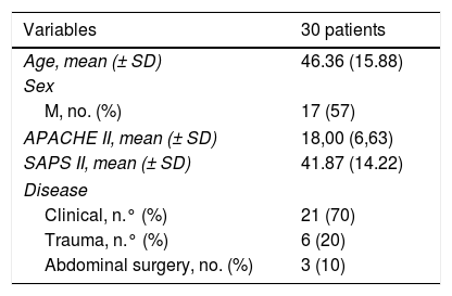
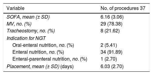
The demographic variables, APACHE II, SAPS II, and disease at ICU admission are shown on Table 1. The details of the variables registered at the moment when the NGT was inserted are shown on Table 2.

Table 1.

Demographic variables, APACHE II, SAPS II, and disease of the patients at the ICU admission.

Variables  30 patients 
Age, mean (± SD)  46.36 (15.88) 
Sex
M, no. (%)  17 (57) 
APACHE II, mean (± SD)  18,00 (6,63) 
SAPS II, mean (± SD)  41.87 (14.22) 
Disease
Clinical, n.° (%)  21 (70) 
Trauma, n.° (%)  6 (20) 
Abdominal surgery, no. (%)  3 (10) 

APACHE II, Acute Physiology and Chronic Health Evaluation II; F, female; ICU, intensive care unit; M, male; SAPS II, Simplified Acute Physiology Score II; SD, standard deviation.

Table 2.

Value of the variables registered at the time when the NGT was inserted.

Variable  No. of procedures 37 
SOFA, mean (± SD)  6.16 (3.06) 
MV, no. (%)  29 (78.38) 
Tracheostomy, no. (%)  8 (21.62) 
Indication for NGT
Oral-enteral nutrition, no. (%)  2 (5.41) 
Enteral nutrition, no. (%)  34 (91.89) 
Enteral-parenteral nutrition, no. (%)  1 (2.70) 
Placement, mean (± SD) (days)  6.03 (2.70) 

MV, mechanical ventilation; NGT, nasogastric tube; SD, standard deviation; SOFA, Sequential Organ Failure Assessment score.

The position of the NGT was correct according to the CDU 32 times and according to the X-ray 31 times. In ambiguous cases, the correct placement of the NGT was identified in the lower third of the esophagus through X-ray. The position was considered incorrect according to both imaging modalities on 5 different occasions.

In this study, the sensitivity, PPV, and NPV estimated to achieve diagnosis on the CDU were 100%, 96.9%, and 100%, respectively. Also, a good correlation was found between the CDU and the X-ray (Spearman’s correlation coefficient, Rho, 0.898; P < .001).

With respect to the necessary time to achieve diagnosis of the correct position of the NGT, the median was 10 min (IQR, 9–15) for the DCU, and 90 min (IQR, 30–120) for the X-ray (P < .001).

Other authors have used ultrasound techniques to identify the NGT inside the gastric antrum with good sensitivity.6–9 Kim et al. used the two-dimensional ultrasound to guide the placement of the NGT and see it run down the cervical esophagus finding a 93% sensitivity and a 98% PPV.10 In approximately half of the cases, additional maneuvers were required to make the imaging modality more sensitive, and a high-resolution ultrasound probe was used for a better visualization of the esophagus, but this is something that may not be reproducible in ICUs with fewer resources.

Zatelli and Vezzali studied ultrasound images in multiple windows (including the instillation of fluid) after NGT insertion without CDU guidance, and they reported an 86.4% sensitivity, an 66,7% specificity, and a 97% PPV.6 Our study found higher sensitivity and specificity rates and a similar predictive value despite the lower number of windows. The advantage here is that the time required is shorter, and that the presence of the doctor is not mandatory during NGT insertion, meaning that it can be inserted by another healthcare professional for later checking. In contrast with the aforementioned study, we chose the instillation of air instead of saline solution due to the possibility of iatrogenesis due to an incorrect NGT insertion in the respiratory tract.

As far as we know, there are no studies on the use of CDU for this purpose. These are some of the limitations of our study we should mention: the small sample size, the populations that were not studied: obesity, pregnancy, hiatal hernia, GI surgery postoperative (because no cases in our sample were found), the use of a single ultrasound window, and the indirect visualization of the nasogastric tube. After the results obtained future studies should be conducted. They should compare the CDU to the two-dimensional mode; sensitivity and specificity changes to the combination of other ultrasound windows; and secondary variations to different velocities during bolus administration.

The CDU proved to be a fast, practical, cost-effective, and highly accurate imaging modality to check the position of the NGT in a small group of adult critically ill patients. Although the CDU may be considered the first-line imaging modality for guidance purposes, studies with larger populations are still needed to confirm these findings.

Funding

This study received no funding whatsoever.

Conflicts of interest

None reported.

Acknowledgements

We wish to thank Dr. Adalberto Baleani, Head of the Imaging Diagnostic Modalities Unit of Hospital Eva Perón.

Appendix A
Supplementary data

The following is Supplementary data to this article:

(0.82MB)

References
[1]
Y. Liu, W. Zhao, W. Chen, X. Shen, R. Fu, Y. Zhao, et al.
Effects of early enteral nutrition on immune function and prognosis of patients with sepsis on mechanical ventilation.
J Intensive Care Med, (2018),
[2]
C. Gubler, P. Bauerfeind, S.R. Vavricka, B. Mullhaupt, M. Fried, S.M. Wildi.
Bedside sonographic control for positioning enteral feeding tubes: a controlled study in intensive care unit patients.
Endoscopy, 38 (2006), pp. 1256-1260
[3]
J.B. Pillai, A. Vegas, S. Brister.
Thoracic complications of nasogastric tube: review of safe practice.
Interact Cardiovasc Thorac Surg, 4 (2005), pp. 429-433
[4]
F. Araimo, R. Caramia, E. Meschesi.
Intracranial placement of a nasogastric tube in a non-trauma patient.
HSR Proc Intensive Care Cardiovasc Anesth, 3 (2011), pp. 71
[5]
V. Rahimi-Movaghar, S.B. Boroojeny, A. Moghtaderi, B. Keshmirian.
Intracranial placement of a nasogastric tube. A lesson to be re-learnt?.
Acta Neurochir (Wien), 147 (2005), pp. 573-574
[6]
M. Zatelli, N. Vezzali.
4-Point ultrasonography to confirm the correct position of the nasogastric tube in 114 critically ill patients.
J Ultrasound, 20 (2017), pp. 53-58
[7]
H. Tsujimoto, Y. Tsujimoto, Y. Nakata, M. Akazawa, Y. Kataoka.
Ultrasonography for confirmation of gastric tube placement.
Cochrane Database Syst Rev, 4 (2017),
[8]
T. Lin, W. Gifford, Y. Lan, X. Qin, X. Liu, J. Wang, et al.
Diagnostic accuracy of ultrasonography for detecting nasogastric tube (NGT) placement in adults: a systematic review and meta analysis.
Int J Nurs Stud, 71 (2017), pp. 80-88
[9]
G. Piton, R. Parel, E. Delabrousse, G. Capellier.
Echography for nasogastric tube placement verification.
Eur J Clin Nutr, 71 (2017), pp. 669-670
[10]
H.M. Kim, B.H. So, W.J. Jeong, S.M. Choi, K.N. Park.
The effectiveness of ultrasonography in verifying the placement of a nasogastric tube in patients with low consciousness at an emergency center.
Scand J Trauma Resusc Emerg Med, 20 (2012), pp. 38

Please cite this article as: Acosta Pedemonte NB, Bagilet DH, Rocchetti NS, Torresan GV, Rodríguez NA, Settecase CJ. La ecografía doppler color es un método preciso para evaluar la posición de la sonda nasogástrica en pacientes críticos. Med Intensiva. 2021;45:e11–e14.

Copyright © 2020. Elsevier España, S.L.U. and SEMICYUC
Download PDF
Idiomas
Medicina Intensiva (English Edition)
Article options
Tools
Supplemental materials
es en

¿Es usted profesional sanitario apto para prescribir o dispensar medicamentos?

Are you a health professional able to prescribe or dispense drugs?